. 2009 Nov 4;101(21):1510-22.
doi: 10.1093/jnci/djp332. Epub 2009 Sep 23.

Association of Merkel cell polyomavirus-specific antibodies with Merkel cell carcinoma

Affiliations

Association of Merkel cell polyomavirus-specific antibodies with Merkel cell carcinoma

Joseph J Carter et al. J Natl Cancer Inst. .

Abstract

Background: Merkel cell polyomavirus (MCPyV) has been detected in approximately 75% of patients with the rare skin cancer Merkel cell carcinoma. We investigated the prevalence of antibodies against MCPyV in the general population and the association between these antibodies and Merkel cell carcinoma.

Methods: Multiplex antibody-binding assays were used to assess levels of antibodies against polyomaviruses in plasma. MCPyV VP1 antibody levels were determined in plasma from 41 patients with Merkel cell carcinoma and 76 matched control subjects. MCPyV DNA was detected in tumor tissue specimens by quantitative polymerase chain reaction. Seroprevalence of polyomavirus-specific antibodies was determined in 451 control subjects. MCPyV strain-specific antibody recognition was investigated by replacing coding sequences from MCPyV strain 350 with those from MCPyV strain w162.

Results: We found that 36 (88%) of 41 patients with Merkel cell carcinoma carried antibodies against VP1 from MCPyV w162 compared with 40 (53%) of the 76 control subjects (odds ratio adjusted for age and sex = 6.6, 95% confidence interval [CI] = 2.3 to 18.8). MCPyV DNA was detectable in 24 (77%) of the 31 Merkel cell carcinoma tumors available, with 22 (92%) of these 24 patients also carrying antibodies against MCPyV. Among 451 control subjects from the general population, prevalence of antibodies against human polyomaviruses was 92% (95% CI = 89% to 94%) for BK virus, 45% (95% CI = 40% to 50%) for JC virus, 98% (95% CI = 96% to 99%) for WU polyomavirus, 90% (95% CI = 87% to 93%) for KI polyomavirus, and 59% (95% CI = 55% to 64%) for MCPyV. Few case patients had reactivity against MCPyV strain 350; however, indistinguishable reactivities were found with VP1 from strain 350 carrying a double mutation (residues 288 and 316) and VP1 from strain w162.

Conclusion: Infection with MCPyV is common in the general population. MCPyV, but not other human polyomaviruses, appears to be associated with Merkel cell carcinoma.


Figures
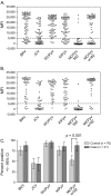
Figure 1
Figure 1
Reactivity against the five human polyomaviruses in serum from 41 case patients with Merkel cell carcinoma and from 76 age- and sex-matched population-based control subjects in control group 1. A) Reactivity of plasma samples from all 76 control subjects in control group 1 against major capsid proteins (VP1 proteins) from five polyomaviruses: BK virus (BKV), JC virus (JCV), WU polyomavirus (WUPyV), KI polyomavirus (KIPyV), and Merkel cell carcinoma polyomavirus (MCPyV strains 350 and w162). Antibody reactivity specific for polyomavirus VP1 proteins was examined by use of a multiplex antibody-binding assay. Each circle represents the median fluorescent intensity (MFI) value from a plasma sample against the fusion protein indicated. B) Reactivity of plasma samples from 41 case patients with Merkel cell carcinoma against VP1 proteins from the five polyomaviruses. These plasma samples were tested as described for the control subjects. C) Percentage of seropositive case patients and control subjects. Percentages of subjects in panels A and B who were seropositive for antibodies against each polyomavirus were determined by use of a cut point for seropositivity of more than 5000 MFI units.
Figure 2
Figure 2
Correlation between reactivity against the major capsid protein (VP1) of Merkel cell polyomavirus (MCPyV) strain w162 and against VP1 proteins of the four other polyomaviruses: BK virus (BKV), JC virus (JCV), WU polyomavirus (WUPyV), and KI polyomavirus (KIPyV). Reactivity was determined in a multiplex antibody-binding assay. Data are expressed as median fluorescent intensity (MFI) for reactivity against VP1 protein of the polyomaviruses as indicated from 76 control subjects from control group 1 and from 41 case patients with Merkel cell carcinoma. No distinctions were made between results from control subjects and from case patients, and each dot represents one plasma sample. Lines = data from linear regression analyses. Correlation coefficients (r2) are at the lower right corners.
Figure 3
Figure 3
Quantitative multiplex binding assay. An antigen-coated bead mixture was incubated with serum that was highly reactive against the major capsid protein (VP1) from MCPyV strain w162 and other polyomaviruses. Before mixing with the antigen-coated beads, the serum was serially diluted 1:5, starting at a dilution of 1:100. Data are the median fluorescent intensity (MFI) values, and the curves were generated by fitting the data to a sigmoidal curve by use of the computer program GraphPad PRISM. BK virus (solid squares), JC virus (open triangles), WU polyomavirus (open circles), KI polyomavirus (open diamonds), Merkel cell polyomavirus strain w162 (solid circles), and Merkel cell polyomavirus strain 350 (open squares). The data are from a representative experiment of two experiments. Results of both experiments were similar.
Figure 4
Figure 4
Identification of amino acid residues in the major capsid protein (VP1) of Merkel cell polyomavirus (MCPyV) 350 that confer low seroreactivity. A) Alignment of VP1 sequences of three MCPyV strains and of the four other human polyomaviruses: BK virus (BKV), JC virus (JCV), KI polyomavirus (KIPyV), and WU polyomavirus (WUPyV). Sequences were obtained from GenBank and aligned with ClustalW (http://align.genome.jp/). Sequences containing residues corresponding to the four divergent residues in the MCPyV VP1 sequence are shown, with divergent residues boxed. Residue numbers refer to those in the MCPyV sequence. GenBank accession numbers for the VP1 sequences shown were FJ392560 for MCPyV w162, EU375803 for MCPyV 350, EU375804 for MCPyV 339, NC_001538 for BKV, NC_001699 for JCV, EF127906 for KIPyV, and EF444549 for WUPyV. B) Identification of VP1 residues essential for antibody recognition of MCPyV strain w162. Point mutations in the MCPyV 350 VP1 sequence and reactivity in plasma against the mutant proteins are shown. Point mutations at positions 288, 316, and 366 were inserted in the MCPyV 350 VP1 sequence either alone or in combination by use of site-directed mutagenesis. All mutant proteins were expressed as GST–VP1 fusion proteins in bacteria and tested in a multiplex antibody-binding assay with 28 plasma samples that had been shown to have reactivity against MCPyV w162 VP1 (Figure 1). The letters listed vertically on the x-axis are the one-letter amino acid abbreviations for residues at the sequence position in the VP1 protein listed on the left. Letters that are white on a black background indicate that the sequence is the same as the MCPyV w162 VP1 sequence and letters that are black on a white background indicate that the sequence is the same as that of MCPyV 350 VP1. Data are the average median fluorescent intensity (MFI) of the 28 plasma samples with reactivity against MCPyV w162 VP1 (one test per sample) after subtracting background and normalizing to the MFI for MCPyV w162. Error bars = 95% confidence intervals. The data are from a representative experiment of an experiment conducted twice. C) Comparison of VP1 sequences from nine MCPyV strains. Partial sequences from seven Merkel cell tumors were compared with the full-length sequences of VP1 proteins from MCPyV strains w162 and MCPyV350 (obtained from GenBank). Sequences around the nonconserved residues 185, 288, 316, and 366 are shown. Immunoreactive amino acids at positions 288 and 316 (shaded) are conserved in all sequences except for that of MCPyV strain 350. The amino acid sequences were identical to those of strains w162 and 339 (data not shown), except for a D221N substitution at position 235 and a stop codon at position 123 in sequence 179 (GenBank accession numbers = FJ649201–FJ649207).
Figure 5
Figure 5
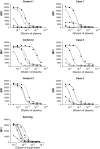
Quantitative measurement of antibodies against the major capsid protein (VP1) of human polyomaviruses. A quantitative multiplex antibody-binding assay was used to measure antibodies in plasma from case patients with Merkel cell carcinoma and control subjects (control group 1) who had tested strongly positive in the multiplex antibody-binding assay (Figure 1). Plasma (or hybridoma supernatant) was diluted to 1:333 followed by eight 1:3 serial dilutions (with the highest dilution = 1:7.29 × 10⁵) and tested for reactivity against VP1 proteins from the following polyomaviruses: BK virus, JC virus, WU polyomavirus, KI polyomavirus, and Merkel cell carcinoma polyomavirus virus (MCPyV). Data from one representative case patient with Merkel cell carcinoma or one representative control subject are shown in each panel. Data from other subjects were similar. Data points are the median fluorescent intensities (MFIs) for each antigen, which is a relative measure of the amount of human IgG bound to the fusion proteins. Curves were generated (and the antibody dilution producing one-half of the maximal binding [EC50 value] was calculated) by fitting the data to an equation for a sigmodal curve. The plasma samples used in this experiment had been identified previously as having high reactivity against the VP1 of MCPyV w162. The data are from a representative experiment of two experiments. Results of both experiments were similar.
Figure 6
Figure 6
Distribution of seroreactivity against the major capsid proteins (VP1) of all five human polyomavirus (BK virus [BKV], JC virus [JCV], WU polyomavirus [WUPyV], KI polyomavirus [KIPyV], and Merkel cell carcinoma polyomavirus virus [MCPyV]) and human papillomavirus (HPV) 16 L1 protein among 451 women in population-based control group 2. The multiplex antibody-binding assay was used to assess seroreactivity in 451 samples previously tested for HPV-16 L1 reactivity (18). The background median fluorescent intensity (MFI) values were subtracted from MFI values for each VP1 and for HPV-16 L1 and data were plotted. Dotted lines indicate the cut points of 15 000 for polyomaviruses and of 820.25 for the HPV-16 L1. The HPV-16 L1 cut point was the highest quartile of values.
Figure 7
Figure 7
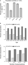
Association of seropositivity against the major capsid protein (VP1) from Merkel cell polyomavirus (MCPyV) w162 or L1 protein from human papillomavirus 16 (HPV-16) with age or number of sexual partners. A) Percentage of women in the general population who are seropositive for polyomaviruses. Reactivity against the major capsid proteins (VP1) of the five human polyomaviruses (BK virus [BKV], JC virus [JCV], WU polyomavirus [WUPyV], KI polyomavirus [KIPyV], and Merkel cell carcinoma polyomavirus virus [MCPyV]) and HPV-16 L1 protein was assessed in serum from 451 control subjects (in the population-based control group 2) by use of the multiplex antibody-binding assay. Data are the proportion of seropositive samples for each polyomavirus type tested. Error bars = 95% confidence intervals (CIs). B) Association of MCPyV and HPV-16 seropositivity with age. Age-stratified seropositivity for HPV-16 (lightly shaded bars) and MCPyV w162 (solid bars). The following numbers of serum were evaluated in each group: 47 in the age group of 18–29 years (MCPyV seropositivity = 55.3%, 95% CI = 40.1% to 69.8%; HPV-16 seropositivity = 42.6%, 95% CI = 28.3% to 57.8%), 122 in the age group of 30–39 years (MCPyV = 55.7%, 95% CI = 46.5% to 64.7%; HPV-16 = 26.2%, 95% CI = 18.7% to 35.0%), 119 in the age group of 40–49 years (MCPyV = 64.7%, 95% CI = 55.4% to 73.2%; HPV-16 = 26.9%, 95% CI = 19.2% to 35.8%), 83 in the age group of 50–59 years (MCPyV = 59.0%, 95% CI = 47.7% to 69.7%; HPV-16 = 16.7%, 95% CI = 9.5% to 26.7%), 46 in the age group of 60–69 years (MCPyV = 65.2%, 95% CI = 49.8% to 78.6%; HPV-16 = 21.7%, 95% CI = 10.9% to 36.4%), and 33 in the age group of 70–74 years (MCPyV = 51.5%, 95% CI = 33.5% to 69.2%; HPV-16 = 12.1%, 95% CI = 3.4% to 28.2%). For the association between seropositivity for HPV-16 L1 and age, Ptrend = .002. For the association between seropositivity for MCPyV and age, Ptrend = .6. C) Association of MCPyV and HPV-16 seropositivity with number of sex partners. Lightly shaded = seropositivity against HPV-16 L1, solid bars = MCPyV w162 VP1. The following numbers of serum samples were evaluated in each group: 123 in the one-partner group (MCPyV seropositivity = 55.7%, 95% CI = 46.5% to 64.7%; HPV-16 seropositivity = 14.8%, 95% CI = 9.0% to 22.3%), 131 in the two- or three-partner group (MCPyV = 61.8%, 95% CI = 52.9% to 70.2%; HPV-16 = 18.3%, 95% CI = 12.1% to 26.0%), 151 in the five- to 14-partner group (MCPyV = 57.6%, 95% CI = 49.3.5% to 65.6%; HPV-16 = 36.4%, 95% CI = 28.8% to 44.6%), and 38 in the group with 15 or more partners (MCPyV = 68.4%, 95% CI = 51.3% to 82.5%; HPV-16 = 39.5%, 95% CI = 24.0% to 56.6%). For the association between seropositivity against HPV-16 L1 and number of sex partners, Ptrend < .001. For the association between seropositivity against MCPyV VP1 and number of sex partners, Ptrend = .5.

References

    1. Poulsen M. Merkel-cell carcinoma of the skin. Lancet Oncol. 2004;5(10):593–599. - PubMed
    1. Heath M, Jaimes N, Lemos B, et al. Clinical characteristics of Merkel cell carcinoma at diagnosis in 195 patients: the AEIOU features. J Am Acad Dermatol. 2008;58(3):375–381. - PMC - PubMed
    1. Feng H, Shuda M, Chang Y, Moore PS. Clonal integration of a polyomavirus in human Merkel cell carcinoma. Science. 2008;319(5866):1096–1100. - PMC - PubMed
    1. Ahuja D, Saenz-Robles MT, Pipas JM. SV40 large T antigen targets multiple cellular pathways to elicit cellular transformation. Oncogene. 2005;24(52):7729–7745. - PubMed
    1. Shuda M, Feng H, Kwun HJ, et al. T antigen mutations are a human tumor-specific signature for Merkel cell polyomavirus. Proc Natl Acad Sci U S A. 2008;105(42):16272–16277. - PMC - PubMed

Publication types

MeSH terms