. 2009 Nov 19;361(21):2046-55.
doi: 10.1056/NEJMoa0905506. Epub 2009 Sep 23.

Combined immunodeficiency associated with DOCK8 mutations

Affiliations

Combined immunodeficiency associated with DOCK8 mutations

Qian Zhang et al. N Engl J Med. .

Abstract

Background: Recurrent sinopulmonary and cutaneous viral infections with elevated serum levels of IgE are features of some variants of combined immunodeficiency. The genetic causes of these variants are unknown.

Methods: We collected longitudinal clinical data on 11 patients from eight families who had recurrent sinopulmonary and cutaneous viral infections. We performed comparative genomic hybridization arrays and targeted gene sequencing. Variants with predicted loss-of-expression mutations were confirmed by means of a quantitative reverse-transcriptase-polymerase-chain-reaction assay and immunoblotting. We evaluated the number and function of lymphocytes with the use of in vitro assays and flow cytometry.

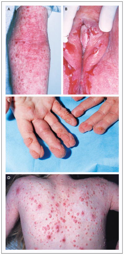
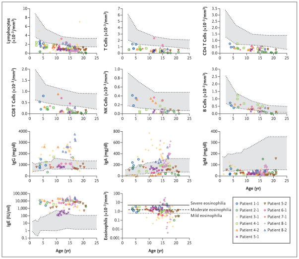
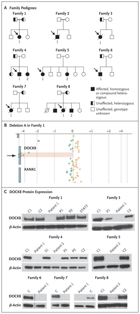
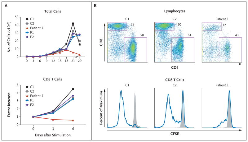
Results: Patients had recurrent otitis media, sinusitis, and pneumonias; recurrent Staphylococcus aureus skin infections with otitis externa; recurrent, severe herpes simplex virus or herpes zoster infections; extensive and persistent infections with molluscum contagiosum; and human papillomavirus infections. Most patients had severe atopy with anaphylaxis; several had squamous-cell carcinomas, and one had T-cell lymphoma-leukemia. Elevated serum IgE levels, hypereosinophilia, low numbers of T cells and B cells, low serum IgM levels, and variable IgG antibody responses were common. Expansion in vitro of activated CD8 T cells was impaired. Novel homozygous or compound heterozygous deletions and point mutations in the gene encoding the dedicator of cytokinesis 8 protein (DOCK8) led to the absence of DOCK8 protein in lymphocytes.

Conclusions: Autosomal recessive DOCK8 deficiency is associated with a novel variant of combined immunodeficiency.


Conflict of interest statement

No potential conflict of interest relevant to this article was reported.

Figures

Figure 1
Figure 1. Characteristic Dermatologic Findings
Among the characteristic dermatologic findings in the patients in the study were atopic dermatitis with scattered superimposed molluscum contagiosum lesions on the inside of the arm (Panel A), chronic ulcerative anogenital herpes simplex virus infection (Panel B), periungual and acral warts (Panel C), and molluscum contagiosum on the back (Panel D).
Figure 2
Figure 2. Immunologic Assessment
Values for blood analyses are shown according to age in the index patients in Families 1, 2, and 3; Patients 1 and 2 in Family 4; Patients 1 and 2 in Family 5; Patient 1 in Family 6; Patient 1 in Family 7; and Patients 1 and 2 in Family 8. In the color key, patients are listed according to their family number, followed by their patient number (Patient 1-1, Patient 2-1, etc.). Normal ranges for age are shown in gray. For lymphocyte subgroups, the normal ranges represent the 10th to 90th percentiles based on published data. For leukocyte subgroups and serum immunoglobulin, the normal ranges are shown with 95% confidence intervals. For eosinophils, the lines represent arbitrary thresholds for mild, moderate, or severe eosinophilia.
Figure 3
Figure 3. Patient Pedigrees and DOCK8 Molecular Analyses
Panel A shows the pedigrees of the eight families in the study. Probands are designated by arrows. Circles represent female family members, and squares male family members. A slash through a symbol represents a deceased person. Panel B shows log2 ratios of DNA from Patient 1 of Family 1 with deletion A (spanning exons 10 through 23) to reference DNA (on the x axis) after hybridization to probes at or near the DOCK8 locus, according to the genomic position (on the y axis). The arrow pointing to the shaded region indicates the homozygous DOCK8 deletion. The green crosses indicate probes where the signal of the log2 ratio was less than zero, and the orange crosses more than zero. Panel C shows immunoblots for DOCK8 protein expression in seven families that were included in the study. Lysates are from primary T cells (for all members of Family 1 and Patient 2 of Family 8), Epstein–Barr virus–transformed B-cell lines (for Families 3, 4, 6, and 7 and Patient 1 of Family 8), and herpesvirus saimiri–transformed T-cell lines (for Family 5). The data for Family 6 show two lanes (samples from a control [C] and from Patient 1) that are aligned from the same gel. Immunoblots for Patient 1 in Family 2 and for Patient 1 in Family 5, which were analyzed after the completion of the study, are shown in Figure 7A in the Supplementary Appendix. No DOCK8 protein was found in samples from any of the 11 patients who were tested. Included in the samples for Family 1 is an unrelated STAT3-mutant control with autosomal dominant hyperimmunoglobulin E syndrome. β-actin is included as a loading control in all the analyses. P denotes parent, and S a heterozygous member of Family 4.
Figure 4
Figure 4. Impaired CD8 T-Cell Activation and Proliferation in Association with DOCK8 Deficiency in Family 1
Panels A and B show the results of two independent experiments in Family 1. In Panel A, after activation of peripheral-blood mononuclear cells (PBMCs) with anti-CD3 plus anti-CD28 antibodies for 3 days, T cells were expanded in culture with interleukin-2. Absolute numbers of CD8 T cells were calculated on the basis of flow cytometric determinations of their percentages; CD8 T-cell numbers over time are shown as factor increases normalized to the prestimulated cell population. In Panel B, PBMCs that were labeled with carboxyfluorescein succinimidyl ester (CFSE) were either unstimulated or stimulated, as described for Panel A. After 3 days, cells were stained with the use of fluorescence-labeled antibodies against T-cell subgroup markers and then analyzed on flow cytometry. The top row shows profiles for stimulated cells, with the location of each dot reflecting an individual cell’s intensity for CD4 staining (on the x axis) versus CD8 staining (on the y axis). The boxes delineate the CD8 and CD4 T-cell subgroups. As compared with control samples, samples from Patient 1 had a lower proportion of CD8 T cells (12% of lymphocytes) after activation. Gated CD8 T cells were further analyzed for the fluorescence intensity of CFSE, which is successively diluted with each cell division. The bottom row shows histograms with the height (y axis) reflecting a percent of maximum of gated CD8 T cells that display any given fluorescence intensity of CFSE (on the x axis). The gray shading indicates unstimulated cells, and the blue line indicates stimulation. C denotes healthy control subject, and P parent.

References

    1. Fischer A. Human primary immunodeficiency diseases. Immunity. 2007;27:835–45. - PubMed
    1. Bustamante J, Zhang SY, von Bernuth H, Abel L, Casanova JL. From infectious diseases to primary immunodeficiencies. Immunol Allergy Clin North Am. 2008;28:235–58. - PubMed
    1. Kawai T, Malech HL. WHIM syndrome: congenital immune deficiency disease. Curr Opin Hematol. 2009;16:20–6. - PMC - PubMed
    1. Prawer SE, Pass F, Vance JC, Greenberg LJ, Yunis EJ, Zelickson AS. Depressed immune function in epidermodysplasia verruciformis. Arch Dermatol. 1977;113:495–9. - PubMed
    1. Renner ED, Puck JM, Holland SM, et al. Autosomal recessive hyperimmunoglobulin E syndrome: a distinct disease entity. J Pediatr. 2004;144:93–9. - PubMed

Publication types

MeSH terms

Substances