

Improving enrichment of circulating fetal DNA for genetic testing: size fractionation followed by whole gene amplification
- PMID: 19776594
- PMCID: PMC2814146
- DOI: 10.1159/000235877
Improving enrichment of circulating fetal DNA for genetic testing: size fractionation followed by whole gene amplification
Abstract
Objective: Among the pitfalls of using cell-free fetal DNA in plasma for prenatal diagnosis is quality of the recovered DNA fragments and concomitant presence of maternal DNA (>95%). Our objective is to provide alternative methods for achieving enrichment and high-quality fetal DNA from plasma.
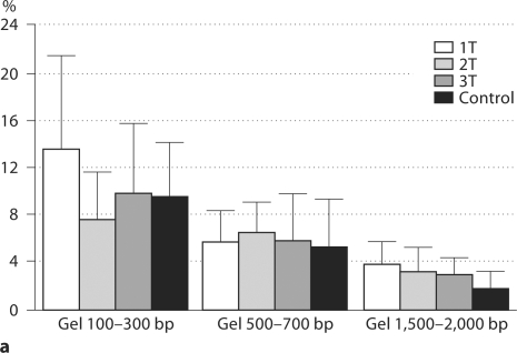
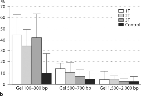
Methods: Cell-free DNA from 31 pregnant women and 18 controls (10 males and 8 females) were size separated using agarose gel electrophoresis. DNA fragments of 100-300, 500-700 and 1,500-2,000 bp were excised and extracted, followed by whole genome amplification (WGA) of recovered fragments. Levels of beta-globin and DYS1 were measured.
Results: Distribution of beta-globin size fragments was similar among pregnant women and controls. Among control male cases, distribution of size fragments was the same for both beta-globin and DYS1. Among maternal cases confirmed to be male, the smallest size fragment (100-300 bp) accounted for nearly 50% (39.76 +/- 17.55%) of the recovered DYS1-DNA (fetal) and only 10% (10.40 +/- 6.49%) of beta-globin (total) DNA. After WGA of plasma fragments from pregnant women, DYS1 sequence amplification was best observed when using the 100-300 bp fragments as template.
Conclusions: Combination of electrophoresis for size separation and WGA led to enriched fetal DNA from plasma. This novel combination of strategies is more likely to permit universal clinical applications of cell-free fetal DNA.
Copyright 2009 S. Karger AG, Basel.
Figures
Similar articles
-
Size fractionation of cell-free DNA in maternal plasma and its application in noninvasive detection of fetal single gene point mutations.Methods Mol Biol. 2008;444:239-51. doi: 10.1007/978-1-59745-066-9_19. Methods Mol Biol. 2008. PMID: 18425486
-
[Non-invasive prenatal genetic diagnosis of beta-thalassaemia using single fetal nucleated erythrocyte from maternal blood].Zhonghua Er Ke Za Zhi. 2007 Dec;45(12):917-21. Zhonghua Er Ke Za Zhi. 2007. PMID: 18339280 Chinese.
-
Microsystem for isolation of fetal DNA from maternal plasma by preparative size separation.Clin Chem. 2009 Dec;55(12):2144-52. doi: 10.1373/clinchem.2009.127480. Epub 2009 Oct 1. Clin Chem. 2009. PMID: 19797718
-
Circulating fetal DNA: its origin and diagnostic potential-a review.Placenta. 2004 Apr;25 Suppl A:S93-S101. doi: 10.1016/j.placenta.2004.01.005. Placenta. 2004. PMID: 15033315 Review.
-
Cell-free fetal nucleic acid testing: a review of the technology and its applications.Obstet Gynecol Surv. 2011 Jul;66(7):431-42. doi: 10.1097/OGX.0b013e31822dfbe2. Obstet Gynecol Surv. 2011. PMID: 21944155 Review.
Cited by
-
Fragment size analysis of free fetal DNA in maternal plasma using Y-STR loci and SRY gene amplification.Nagoya J Med Sci. 2011 Aug;73(3-4):129-35. Nagoya J Med Sci. 2011. PMID: 21928694 Free PMC article.
-
Genome-Wide Noninvasive Prenatal Diagnosis of De Novo Mutations.Methods Mol Biol. 2021;2243:249-269. doi: 10.1007/978-1-0716-1103-6_12. Methods Mol Biol. 2021. PMID: 33606261
-
Epigenetic biomarkers: Current strategies and future challenges for their use in the clinical laboratory.Crit Rev Clin Lab Sci. 2017 Nov-Dec;54(7-8):529-550. doi: 10.1080/10408363.2017.1410520. Epub 2017 Dec 11. Crit Rev Clin Lab Sci. 2017. PMID: 29226748 Free PMC article. Review.
-
Cell-free DNA for genomic profiling and minimal residual disease monitoring in Myeloma- are we there yet?Am J Blood Res. 2020 Jun 15;10(3):26-45. eCollection 2020. Am J Blood Res. 2020. PMID: 32685257 Free PMC article. Review.
-
Automated size selection for short cell-free DNA fragments enriches for circulating tumor DNA and improves error correction during next generation sequencing.PLoS One. 2018 Jul 25;13(7):e0197333. doi: 10.1371/journal.pone.0197333. eCollection 2018. PLoS One. 2018. PMID: 30044795 Free PMC article.
References
-
- Bischoff FZ, Nguyen DD, Marquez-Do D, Moise KJ, Jr, Simpson JL, Elias S. Noninvasive determination of fetal RhD status using fetal DNA in maternal serum and PCR. J Soc Gynecol Investig. 1999;6:64–69. - PubMed
-
- Bischoff FZ, Sinacori MK, Dang DD, Marquez-Do D, Horne C, Lewis DE, Simpson JL. Cell-free fetal DNA and intact fetal cells in maternal blood circulation: implications for first and second trimester non-invasive prenatal diagnosis. Hum Reprod Update. 2002;8:493–500. - PubMed
-
- Bischoff FZ, Dang DX, Marquez-Do D, Martinez D, Horne C, Lewis DE, Simpson JL. Detecting fetal DNA from dried maternal blood spots: another step towards broad scale non-invasive prenatal genetic screening and feasible testing. Reprod Biomed Online. 2003;6:349–351. - PubMed
-
- Johnson KL, Dukes KA, Vidaver J, LeShane ES, Ramirez I, Weber WD, Bischoff FZ, Hahn S, Sharma A, Dang DX, Hire LM, Bianchi DW, Simpson JL, Holzgreve W, Elias S, Klinger KW. Interlaboratory comparison of fetal male DNA detection from common maternal plasma samples by real-time PCR. Clin Chem. 2004;50:516–521. - PubMed
Publication types
MeSH terms
Substances
Grants and funding
LinkOut - more resources
Full Text Sources
Other Literature Sources
Medical
