. 2009 Sep 24;4(9):e7158.
doi: 10.1371/journal.pone.0007158.

miR-155 inhibition sensitizes CD4+ Th cells for TREG mediated suppression

Affiliations

miR-155 inhibition sensitizes CD4+ Th cells for TREG mediated suppression

Heiko F Stahl et al. PLoS One. .

Abstract

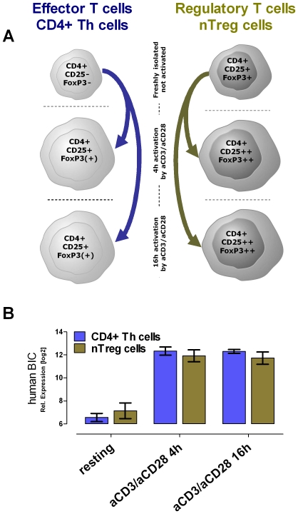
Background: In humans and mice naturally occurring CD4(+)CD25(+) regulatory T cells (nTregs) are a thymus-derived subset of T cells, crucial for the maintenance of peripheral tolerance by controlling not only potentially autoreactive T cells but virtually all cells of the adaptive and innate immune system. Recent work using Dicer-deficient mice irrevocably demonstrated the importance of miRNAs for nTreg cell-mediated tolerance.

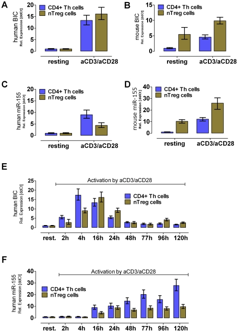
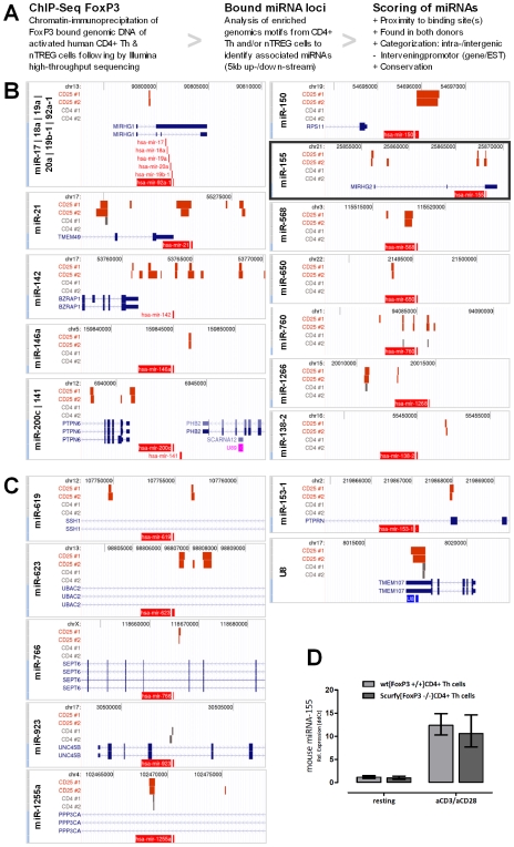
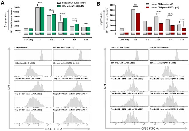
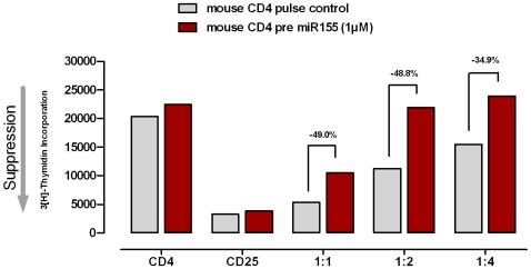
Principal findings: DNA-Microarray analyses of human as well as murine conventional CD4(+) Th cells and nTregs revealed a strong up-regulation of mature miR-155 (microRNA-155) upon activation in both populations. Studying miR-155 expression in FoxP3-deficient scurfy mice and performing FoxP3 ChIP-Seq experiments using activated human T lymphocytes, we show that the expression and maturation of miR-155 seem to be not necessarily regulated by FoxP3. In order to address the functional relevance of elevated miR-155 levels, we transfected miR-155 inhibitors or mature miR-155 RNAs into freshly-isolated human and mouse primary CD4(+) Th cells and nTregs and investigated the resulting phenotype in nTreg suppression assays. Whereas miR-155 inhibition in conventional CD4(+) Th cells strengthened nTreg cell-mediated suppression, overexpression of mature miR-155 rendered these cells unresponsive to nTreg cell-mediated suppression.

Conclusion: Investigation of FoxP3 downstream targets, certainly of bound and regulated miRNAs revealed the associated function between the master regulator FoxP3 and miRNAs as regulators itself. miR-155 is shown to be crucially involved in nTreg cell mediated tolerance by regulating the susceptibility of conventional human as well as murine CD4(+) Th cells to nTreg cell-mediated suppression.


Conflict of interest statement

Competing Interests: HFS, TF, WR, AW, MCL, DM are employees of Boehringer Ingelheim Pharma GmbH & CoKG. PL and VA are employees of Genpathway Inc. Both companies, Boehringer Ingelheim Pharma GmbH & CoKG and Genpathway Inc., were not involved in the decision to publish or preparation of the manuscript.

Figures

Figure 1
Figure 1. Human Exon Array Genechip expression profiling showed BIC as one of the highest up-regulated genes after T cell activation.
(A) Schematic overview of different T cell populations out of 10 human donors. Expression profiles were analyzed of freshly isolated resting CD4+ Th cells and of nTregs. In addition, both populations were profiled upon 4 h and 16 h anti-CD3/anti-CD28 stimulation. (B) The expression profiling revealed the BIC transcript specifically up-regulated upon activation in both populations: the CD4+ Th cells and nTregs. The median relative expression level of BIC in logarithmic scale and the standard deviation is shown (n = 10).
Figure 2
Figure 2. RT-PCR expression analysis of pre-mature BIC transcript and its processed microRNA miR-155 in mice and men.
The BIC transcript is strongly up-regulated upon activation using anti-CD3/anti-CD28 mAb in human donors (A) and in C57/BL6 mice (B). Whereas, in C57/BL6 mice the BIC transcript as well as the matured miR-155 was found to be higher expressed in nTregs than in CD4+ Th cells (B) and (D) (n = 3). In human cells, BIC and the matured form miR-155 were not present in resting cells, but strongly elevated levels were found in Th cells as well as in nTreg cells after TCR activation (A) and (C) (n = 4). Analyzing the kinetic of BIC/miR-155 expression RNA was collected using an activation time course experiment. (E) The activation of human T cell populations showed a temporary activation of the BIC transcription. Whereas, the CD4+ Th cells reached their maximum after 4 h upon activation, the peak for nTreg cells is shifted to the 16 h time point. (F) The levels of matured miR-155 were found to permanently increase within time (until 120 h) in activated CD4+ Th cells, whereas in nTregs a plateau was reached after 24 h. All values were calculated as relative fold changes using the ddCT method. As normalizer RNA Pol II (human & mouse BIC) as well as U18 (human miR-155) and 5S (mouse miR-155) were used.
Figure 3
Figure 3. BIC/miR-155 expression is not necessarily regulated by FoxP3.
(A) Schematic overview of the ChIP-Seq analysis workflow. Genomic loci of all significantly and reproducible FoxP3-bound micro-RNAs are shown: intragenic micro-RNA (B) and intergenic located micro-RNAs (C). Each genomic capture shows in light red the micro-RNA(s) and in blue the overlapping annotated gene(s) including the intro/exon structure(s). In dark red (CD25+/nTREG) and in gray (CD4+ Th cell) the FoxP3-bound genomic regions of both donors are symbolized. In addition, every capture contains the underlying chromosome including the basepair coordinates. The visualisations were generated using the UCSC genome browser (human genome assembly of March 2006). (D) Using Taqman RT-PCR, the expression analysis of T lymphocytes for mature miR-155 showed no significant difference between wild type C57/BL6 and FoxP3 mutated Scurfy mice. Post activation (19 h) an increased expression of matured miR-155 was detectable, in both CD4+ Th cells of wild type and Scurfy mice. All values were calculated as relative fold changes using the ddCT method. As normalizer 5S was used (n = 3).
Figure 4
Figure 4. Modulation of miR-155 levels in CD4+ Th cells influenced the susceptibility to nTreg-mediated suppression.
(A) Blocking the biological available miR-155 by transfection of synthetic anti-miR-155 molecules led to an increased sensibility for nTreg-mediated suppression. Depending on the ratio of CD4+ Th cells to nTregs (4∶1 and 8∶1) a nearly 25% increased susceptibility for suppression of proliferation was observed. The CD4+ Th cell population alone showed no change in proliferation rate in comparison to the pulsed only control population. (B) Increasing the miR-155 levels within CD4+ Th cells by transfection of synthetic miR-155 decrease the susceptibility for nTreg-mediated suppression measured in CFSE proliferation assays. Parallel assay setup revealed a nearly 25% decreased sensibility for suppression of proliferation. The CD4+ Th cell population alone showed also an elevated proliferation in comparison to the control-miRNA transfected population. Shown are the MFI (mean fluorescence intensity) of CFSE labelled CD4+ Th cells after four days of activation (aCD3 and APC). The bar plot diagrams are indicating the grade of suppression levels. The added numbers are showing the differences (%) for the susceptibility of nTreg-mediated suppression of proliferation of the activated CD4+ Th cells.
Figure 5
Figure 5. Overexpression of mature miR-155 rendered murine CD4+ Th cells unresponsive to nTreg-mediated suppression.
Suppression assay performed with naïve murine CD4+ Th cells and murine nTregs: Th cells transfected with pre miR-155 showed up to 40% decreased susceptibility for nTreg cell-mediated suppression compared to the control-transfected CD4+ Th cells. Shown is the 3H-thymidine uptake after 18 h. The bar plot diagrams are indicating the grade of suppression levels. The added numbers are showing the differences (%) for the susceptibility of nTreg-mediated suppression of proliferation of the activated CD4+ Th cells. One representative experiment out of at least three experiments is shown.

References

    1. Liston A, Lu LF, O'Carroll D, Tarakhovsky A, Rudensky AY. Dicer-dependent microRNA pathway safeguards regulatory T cell function. J Exp Med. 2008;205:1993–2004. - PMC - PubMed
    1. Muljo SA, Ansel KM, Kanellopoulou C, Livingston DM, Rao A, et al. Aberrant T cell differentiation in the absence of Dicer. J Exp Med. 2005;202:261–269. - PMC - PubMed
    1. Zhou X, Jeker LT, Fife BT, Zhu S, Anderson MS, et al. Selective miRNA disruption in T reg cells leads to uncontrolled autoimmunity. J Exp Med. 2008;205:1983–1991. - PMC - PubMed
    1. Clurman BE, Hayward WS. Multiple proto-oncogene activations in avian leukosis virus-induced lymphomas: evidence for stage-specific events. Mol Cell Biol. 1989;9:2657–2664. - PMC - PubMed
    1. Eis PS, Tam W, Sun L, Chadburn A, Li Z, et al. Accumulation of miR-155 and BIC RNA in human B cell lymphomas. Proc Natl Acad Sci U S A. 2005;102:3627–3632. - PMC - PubMed

Publication types