. 2009 Sep 24;4(9):e7174.
doi: 10.1371/journal.pone.0007174.

Lymphocyte display: a novel antibody selection platform based on T cell activation

Affiliations

Lymphocyte display: a novel antibody selection platform based on T cell activation

Vanesa Alonso-Camino et al. PLoS One. .

Abstract

Since their onset, display technologies have proven useful for the selection of antibodies against a variety of targets; however, most of the antibodies selected with the currently available platforms need to be further modified for their use in humans, and are restricted to accessible antigens. Furthermore, these platforms are not well suited for in vivo selections. We present here a novel cell based antibody display platform, which takes advantage of the functional capabilities of T lymphocytes. The display of antibodies on the surface of T lymphocytes, as a part of a chimeric-immune receptor (CIR) mediating signaling, may ideally link the antigen-antibody interaction to a demonstrable change in T cell phenotype, due to subsequent expression of the early T cell activation marker CD69. In this proof-of-concept, an in vitro selection was carried out using a human T cell line lentiviral-transduced to express a tumor-specific CIR on the surface, against a human tumor cell line expressing the carcinoembryonic antigen. Based on an effective interaction between the CIR and the tumor antigen, we demonstrated that combining CIR-mediated activation with FACS sorting of CD69(+) T cells, it is possible to isolate binders to tumor specific cell surface antigen, with an enrichment factor of at least 10(3)-fold after two rounds, resulting in a homogeneous population of T cells expressing tumor-specific CIRs.


Conflict of interest statement

Competing Interests: The authors have declared that no competing interests exist.

Figures

Figure 1
Figure 1. Schematic representation of lentiviral vector constructs.
(A) Control monocistronic vector (pRRL-IRES-EGFP) containing only the enhanced-green fluorescent protein (EGFP) gene and bicistronic vector (pRRL-αCEA-CIR-IRES-EGFP) containing the chimeric immune receptor (CIR) gene and the EGFP sequence. LTR, long terminal repeats; ΔGAG, ATG-deleted group specific antigen; RRE, Rev-responsive cis-acting element; CMV promoter; ECMV IRES, enceohalomyocarditis virus internal ribosomal entry site. (B) FACS analysis of EGFP expression after transduction of Jurkat cells with EGFP encoding lentiviral vectors at different MOIs, ranging from 0.25 to 10. (C) FACS analysis of EGFP and cell surface CIR expression after transduction of Jurkat cells with CIR-EGFP encoding lentiviral vectors at MOI of 1.
Figure 2
Figure 2. IL-2 production by JurkatEGFP and JurkatαCEA-CIR-EGFP cells stimulated either with plastic immobilized anti-CD3 mAb or target cells (E∶T = 1∶1; HeLa or HeLaCEA) for 48 hours.
Figure 3
Figure 3. CIR-mediated activation of human T cells.
(A) Cell-surface expression of CEACAM5 (CEA) and NIP-modified molecules on HeLa, HeLaCEA and HeLa cells labeled with 2.5 µg/mL of the hapten (HeLaNIP). (B) FACS analysis of CD69 expression by JurkatEGFP, JurkatαCEA-CIR-EGFP and JurkatαNIP-CIR stimulated either with immobilized anti-CD3 mAb or target cells (E∶T = 1∶1; HeLa, HeLaCEA or HeLaNIP) for 16 hours.
Figure 4
Figure 4. CIR-mediated activation of human T cells.
(A) Cell-surface expression of CEACAM5 (CEA) on HeLa, HT1080, MDA-MB-231 and MKN45 cells. (B) FACS analysis of CD69 expression by Jurkat, JurkatαCEA-CIR-EGFP and JurkatαNIP-CIR stimulated either with immobilized anti-CD3 mAb or target cells (E∶T = 1∶1; HeLa, HT1080, MDA-MB-231 or MKN45) for 16 hours.
Figure 5
Figure 5. Effect of competitor populations on CIR-mediated T cell activation.
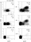
CIR-positive JurkatαCEA-CIR-EGFP effector (E) cells were stimulated with CEA-positive target cells for 16 hours in the presence of increasing amounts of CIR-negative Jurkat competitor (C) cells. The expression of CD69 on E∶C ratios ranging from 100∶1 to 1∶100 was measured by FACS on pre-activation (left panels) and post-activation (right panels) mixtures.
Figure 6
Figure 6. Selection of CIR-activated T cells.
JurkatαCEA-CIR-EGFP effector (E) cells and CIR-negative Jurkat competitor (C) cells at a E∶C mixing ratio 1∶1000 were stimulated with CEA-positive target cells for 16 hours and further sorted on the basis of EGFP and CD69 expression. After a period of cell expansion the activation/selection cycle was repeated.
Figure 7
Figure 7. Selection of CIR-activated T cells.
JurkatαCEA-CIR-EGFP effector (E) cells and CIR-positive JurkatαNIP-CIR competitor (C) cells at a E∶C mixing ratio 1∶1000 were stimulated with CEA-positive target cells for 16 hours and further sorted on the basis of EGFP and CD69 expression. After a period of cell expansion the activation/selection cycle was repeated.
Figure 8
Figure 8. Phenotypic and functional characterization of selected JurkatαCEA-CIR-EGFP cells.
(A) Comparative analysis of EGFP and cell surface CIR expression of the initial (JurkatαCEA-CIR-EGFP) and the selected (JurkatαCEA-CIR-EGFP/2S) cell population after two rounds of activation/selection on CEA-positive target cells. (B) Comparative analysis of CD69 expression by JurkatαCEA-CIR-EGFP and JurkatαCEA-CIR-EGFP/2S cell populations after stimulation either with immobilized anti-CD3 mAb or target cells (E∶T = 1∶1; HeLa or HeLaCEA).

References

    1. Smith GP. Filamentous fusion phage: novel expression vectors that display cloned antigens on the virion surface. Science. 1985;14;228 (4705):1315–1317. - PubMed
    1. Winter G, Griffiths AD, Hawkins RE, Hoogenboom HR. Making antibodies by phage display technology. Annu Rev Immunol. 1994;12:433–455. - PubMed
    1. Urban JH, Schneider RM, Compte M, Finger C, Cichutek K, et al. Selection of functional human antibodies from retroviral display libraries. Nucleic Acids Res. 2005;33:e35. - PMC - PubMed
    1. Hoogenboom HR. Selecting and screening recombinant antibody libraries. Nat Biotechnol. 2005;23:1105–1116. - PubMed
    1. Buchholz CJ, Peng KW, Morling FJ, Zhang J, Cosset FL, et al. In vivo selection of protease cleavage sites from retrovirus display libraries. Nat Biotechnol. 1998;16:951–954. - PubMed

Publication types