. 2010 Feb;34(2):263-9.
doi: 10.1007/s00264-009-0873-3. Epub 2009 Sep 24.

Osteotomy around young deformed knees: 38-year super-long-term follow-up to detect osteoarthritis

Affiliations

Osteotomy around young deformed knees: 38-year super-long-term follow-up to detect osteoarthritis

Tomihisa Koshino. Int Orthop. 2010 Feb.

Abstract

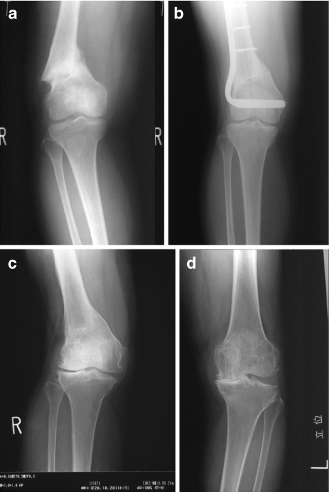
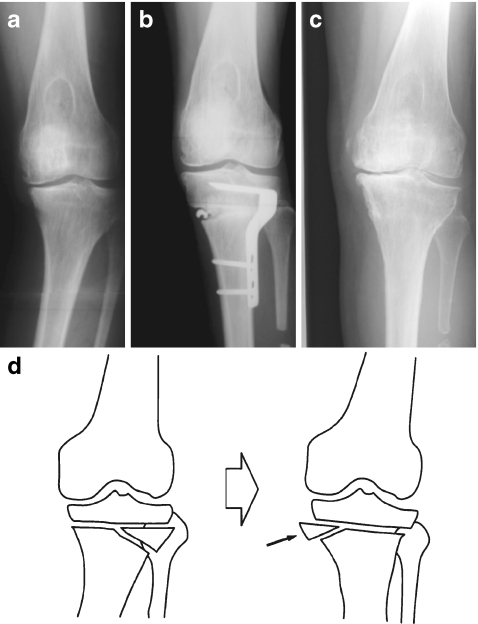
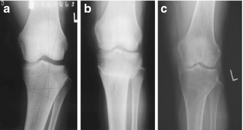
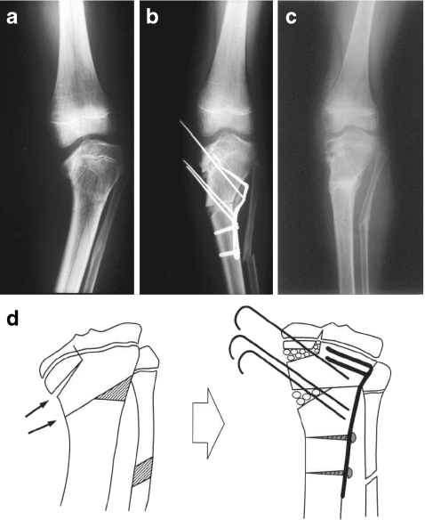
Since 1969 corrective osteotomy has been performed at our institute in young patients (under 40 years) with bowlegs, knock knees and flexion or rotational deformities around the knee. Fifty-seven knees (29 left, 28 right) of 45 patients (19 boys, 26 girls) were followed-up for a period ranging from 30 to 38 years in seven patients with seven knees, from 20 to 29 years in nine patients with 11 knees, and from ten to 19 years in 29 patients with 39 knees. Supracondylar femoral osteotomy was performed on 12 knees (11 patients), high tibial osteotomy above the tibial tuberosity on eight knees (six patients) and below the tuberosity on 37 knees (28 patients). At the final follow-up (age range 42-73 years), all of the deformities were satisfactorily corrected, with no symptoms apart from nine knees, seven of which had dull pain after strenuous sport with osteophytes, etc. in the radiograph. Total knee arthroplasty was performed in the remaining two knees, at ten and 26 years, respectively, after the initial osteotomy. Osteoarthritis developed in the contralateral knee to the initial osteotomy in two patients after 34 years at age 73 and after 33 years at age 67.


Figures

Fig. 1
Fig. 1
Malunited supracondylar fracture of right femur with varus angulation (a). This female patient sustained this fracture at age 25 years. Supracondylar osteotomy of the femur was performed with resection of a medially-based wedge and fixation using an L-shaped blade plate from the lateral side at age 34 years (b). The patient has had no symptoms and no problems. At age 67 years, 33 years after the initial osteotomy, recent final follow-up revealed no pain in the right knee with range of motion from 0° extension to 145° flexion, though osteosclerosis was observed around the lateral femoro-tibial articulation with a femoro-tibial angle of 165° (15° anatomical valgus angulation) in the standing radiograph (c). However, she developed painful osteoarthritis in her left knee with obliteration of the medial joint space and varus deformity in the standing radiograph (d)
Fig. 2
Fig. 2
Varus deformity after osteomyelitis of left femur at age 2 years (a). Hemi-open wedge high tibial osteotomy was performed and the fragments were fixed with a Koshino blade plate laterally, together with segmental resection of the fibular shaft (b) at age 28 years. The resected fibular shaft was cut longitudinally and embedded into the medial opening space of the osteotomy (b). The patient has had no symptoms or problems. Recent final follow-up revealed no pain in the left knee, with range of knee motion from 0° extension to 120° flexion. The standing radiograph taken at age 59 years, 31 years after the initial osteotomy, showed satisfactory limb alignment with a femoro-tibial angle of 171° (9° anatomical valgus angulation) (c). As for hemi-open wedge high tibial osteotomy, the horizontal line of osteotomy was marked out 20 mm below the joint line, from the mid point of which a small laterally-based wedge was marked out laterally. After removal of the wedge the lateral side of the osteotomy site was closed and bone grafts were embedded into the medial opened space (d)
Fig. 3
Fig. 3
Varus deformity of left knee due to rickets with standing femoro-tibial angle of 194° (14° anatomical varus angulation in a 21-year-old woman) (a). Tibial osteotomy was performed to remove a 24° laterally-based wedge just below the tibial tuberosity and the fragments were fixed with crossed Kirschner wires and a long leg plaster splint. The K-wires were drawn out one by one every week from 3 weeks after osteotomy and a long leg cylinder cast was applied. The standing radiograph one year after osteotomy showed a femoro-tibial angle of 170° (10° of anatomical valgus angulation) (b). The patient has been doing well with no problems. Recent final follow-up at age 59 years, 38 years after the initial osteotomy, revealed no pain in the left knee with full range of motion and the standing radiograph showed a femoro-tibial angle of 170° (c)
Fig. 4
Fig. 4
Severe varus deformity of left knee due to Blount’s disease (a). The preoperative standing radiograph showed depression of the medial proximal epiphysis of the tibia with a femoro-tibial angle of 201° (21° anatomical varus angulation). Trans-epiphyseal opening wedge osteotomy together with hemi-open wedge osteotomy in the region below the tibial tuberosity was performed at age 8 years, the fragments of which were fixed with oblique Kirschner wires and a hook plate laterally (b). The postoperative standing radiograph 1 year after the initial osteotomies showed a femoro-tibial angle of 169° (11° anatomical valgus angulation). As for trans-epiphyseal opening wedge osteotomy, transverse osteotomy was performed at a level 1 cm below the medial epiphyseal plate and extended upward vertically around the central portion of the metaphysis through the epiphyseal line (d). Bone grafts were embedded into the opened osteotomised space. The fragments were fixed with obliquely inserted Kirschner wires

References

    1. Aglietti P, Rinonapoli E, Stringa G, et al. Tibial osteotomy for the varus osteoarthritic knee. Clin Orthop. 1983;176:239–251. - PubMed
    1. Akizuki S, Shibakawa A, Takizawa T, et al. The long-term outcome of high tibial osteotomy. A ten to 20 year follow up. J Bone Joint Surg. 2008;90-B:592–596. doi: 10.1302/0301-620X.90B5.20386. - DOI - PubMed
    1. Bauer GCH, Insall JN, Koshino T. Tibial osteotomy in gonarthrosis (osteoarthritis of the knee) J Bone Joint Surg. 1969;51-A:1545–1563. - PubMed
    1. Billings A, Scott DF, Camargo MP, et al. High tibial osteotomy with a calibrated osteotomy guide, rigid internal fixation, and early motion. Long-term follow-up. J Bone Joint Surg. 2000;82-A:70–79. - PubMed
    1. Coventry MB, Ilstrup DM, Wallrich SL. Proximal tibial osteotomy – a critical long-term study of eighty-seven cases. J Bone Joint Surg. 1993;75A:196–201. - PubMed

MeSH terms