

Central nesfatin-1-expressing neurons are sensitive to peripheral inflammatory stimulus
- PMID: 19778412
- PMCID: PMC2762958
- DOI: 10.1186/1742-2094-6-27
Central nesfatin-1-expressing neurons are sensitive to peripheral inflammatory stimulus
Abstract
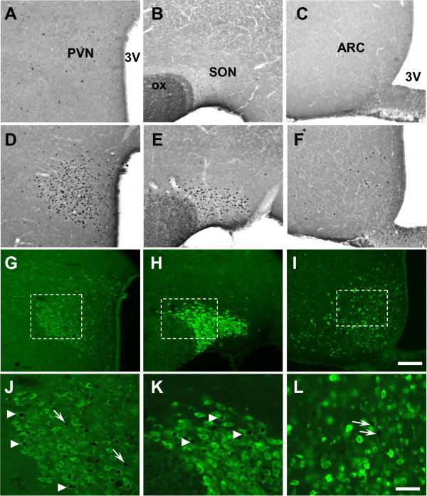
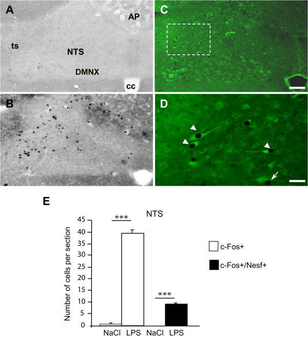
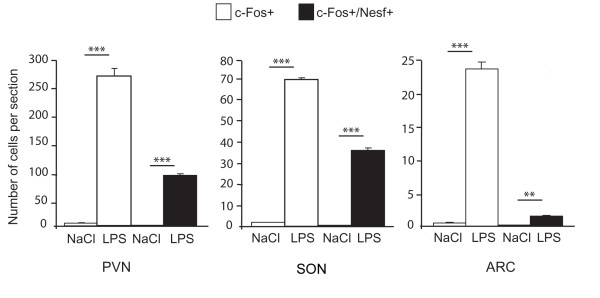
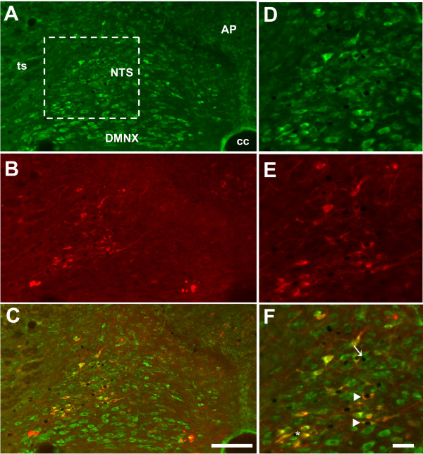
Recently, a novel factor with anorexigenic properties was identified and called nesfatin-1. This protein (82 aac) is not only expressed in peripheral organs but it is also found in neurons located in specific structures including the hypothalamus and the brainstem, two sites strongly involved in food intake regulation. Here, we studied whether some of the neurons that become activated following an injection of an anorectic dose of lipopolysaccharides (LPS) exhibit a nesfatin-1 phenotype. To this end, we used double immunohistochemistry to target the expression of the immediate-early gene c-fos and of nesfatin-1 on coronal frozen sections of the rat brain. The number of c-Fos+/nesfatin-1+ neurons was evaluated in the immunosensitive structures reported to contain nesfatin-1 neurons; i.e. paraventricular hypothalamic nucleus (PVN), supraoptic nucleus (SON), arcuate nucleus (ARC) and nucleus of the solitary tract (NTS). LPS strongly increased the number of c-Fos+/nesfatin-1+ neurons in the PVN, SON and NTS, and to a lesser extent in the ARC. Triple labeling showed that a portion of the nesfatin-1 neurons activated in response to LPS within the NTS are catecholaminergic since they co-express tyrosine hydroxylase (TH). Our data therefore indicate that a portion of nesfatin-1 neurons of both the hypothalamus and brainstem are sensitive to peripheral inflammatory signals, and provide the first clues suggesting that centrally released nesfatin-1 may contribute to the neural mechanisms leading to endotoxaemic anorexia.
Figures
Similar articles
-
Abdominal surgery activates nesfatin-1 immunoreactive brain nuclei in rats.Peptides. 2010 Feb;31(2):263-70. doi: 10.1016/j.peptides.2009.11.015. Epub 2009 Nov 26. Peptides. 2010. PMID: 19944727 Free PMC article.
-
Peripheral injection of bombesin induces c-Fos in NUCB2/nesfatin-1 neurons.Brain Res. 2016 Oct 1;1648(Pt A):46-53. doi: 10.1016/j.brainres.2016.07.006. Epub 2016 Jul 7. Brain Res. 2016. PMID: 27396908
-
Activation of Nesfatin-1-Containing Neurones in the Hypothalamus and Brainstem by Peripheral Administration of Anorectic Hormones and Suppression of Feeding via Central Nesfatin-1 in Rats.J Neuroendocrinol. 2016 Sep;28(9). doi: 10.1111/jne.12400. J Neuroendocrinol. 2016. PMID: 27203571
-
A new anorexigenic protein, nesfatin-1.Peptides. 2009 May;30(5):995-8. doi: 10.1016/j.peptides.2009.01.002. Peptides. 2009. PMID: 19452636 Review.
-
The role of nesfatin-1 in kidney diseases.Pediatr Nephrol. 2025 Apr;40(4):901-907. doi: 10.1007/s00467-024-06569-1. Epub 2024 Oct 31. Pediatr Nephrol. 2025. PMID: 39480586 Free PMC article. Review.
Cited by
-
Electroacupuncture negatively regulates the Nesfatin-1/ERK/CREB pathway to alleviate HPA axis hyperactivity and anxiety-like behaviors caused by surgical trauma.Chin Med. 2024 Aug 17;19(1):108. doi: 10.1186/s13020-024-00974-2. Chin Med. 2024. PMID: 39153974 Free PMC article.
-
Nesfatin-1 Improve Spatial Memory Impairment Following Transient Global Cerebral Ischemia/Reperfusion via Inhibiting Microglial and Caspase-3 Activation.J Mol Neurosci. 2018 Jul;65(3):377-384. doi: 10.1007/s12031-018-1105-3. Epub 2018 Jun 28. J Mol Neurosci. 2018. PMID: 29956089
-
Nesfatin-1--role as possible new potent regulator of food intake.Regul Pept. 2010 Aug 9;163(1-3):18-23. doi: 10.1016/j.regpep.2010.05.002. Epub 2010 May 16. Regul Pept. 2010. PMID: 20580651 Free PMC article. Review.
-
Nesfatin-1: a novel inhibitory regulator of food intake and body weight.Obes Rev. 2011 Apr;12(4):261-71. doi: 10.1111/j.1467-789X.2010.00770.x. Obes Rev. 2011. PMID: 20546141 Free PMC article. Review.
-
Lipopolysaccharide increases gastric and circulating NUCB2/nesfatin-1 concentrations in rats.Peptides. 2011 Sep;32(9):1942-7. doi: 10.1016/j.peptides.2011.07.006. Epub 2011 Jul 18. Peptides. 2011. PMID: 21782869 Free PMC article.
References
-
- Dantzer R. Cytokine-induced sickness behavior: mechanisms and implications. Ann N Y Acad Sci. 2001;933:222–34. - PubMed
Publication types
MeSH terms
Substances
LinkOut - more resources
Full Text Sources
