. 2009 Sep 24:8:76.
doi: 10.1186/1476-4598-8-76.

Blockade of Wnt-1 signaling leads to anti-tumor effects in hepatocellular carcinoma cells

Affiliations

Blockade of Wnt-1 signaling leads to anti-tumor effects in hepatocellular carcinoma cells

Wei Wei et al. Mol Cancer. .

Abstract

Background: Hepatocellular carcinoma (HCC) is an aggressive cancer, and is the third leading cause of cancer death worldwide. Standard therapy is ineffective partly because HCC is intrinsically resistant to conventional chemotherapy. Its poor prognosis and limited treatment options make it critical to develop novel and selective chemotherapeutic agents. Since the Wnt/beta-catenin pathway is essential in HCC carcinogenesis, we studied the inhibition of Wnt-1-mediated signaling as a potential molecular target in HCC.

Results: We demonstrated that Wnt-1 is highly expressed in human hepatoma cell lines and a subgroup of human HCC tissues compared to paired adjacent non-tumor tissues. An anti-Wnt-1 antibody dose-dependently decreased viability and proliferation of Huh7 and Hep40 cells over-expressing Wnt-1 and harboring wild type beta-catenin, but did not affect normal hepatocytes with undetectable Wnt-1 expression. Apoptosis was also observed in Huh7 and Hep40 cells after treatment with anti-Wnt-1 antibody. In these two cell lines, the anti-Wnt-1 antibody decreased beta-catenin/Tcf4 transcriptional activities, which were associated with down-regulation of the endogenous beta-catenin/Tcf4 target genes c-Myc, cyclin D1, and survivin. Intratumoral injection of anti-Wnt-1 antibody suppressed in vivo tumor growth in a Huh7 xenograft model, which was also associated with apoptosis and reduced c-Myc, cyclin D1, and survivin expressions.

Conclusion: Our results suggest that Wnt-1 is a survival factor for HCC cells, and that the blockade of Wnt-1-mediated signaling may offer a potential pathway-specific therapeutic strategy for the treatment of a subgroup of HCC that over-expresses Wnt-1.


Figures

Figure 1
Figure 1
Expression of Wnt-1 protein in human HCC tumors and cell lines. A). Western blot detection of Wnt-1 expression in human HCC tumor tissues (T) and the corresponding adjacent non-tumor liver tissues (N). B). Western blot detection of Wnt-1 expression in HCC cell lines (HepG2, Huh7, and Hep40), and normal hepatocytes from three donors (Hu0910, Hu4122, Hu4074). The immunoblots were quantified by densitometry and the intensities normalized with those of β-actin and given beneath each band.
Figure 2
Figure 2
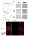
In vitro anti-proliferative and apoptotic effects of anti-Wnt-1 antibody on human HCC cell lines. A). Cell viability assays based on cellular ATP content were used to determine the effect of anti-Wnt-1 antibody on three human HCC cell lines and normal hepatocytes from three donors following 72 hr of antibody treatment. Relative ATP activity is proportional to the number of viable cells. Three independent experiments were done, each in triplicates. B). Phase-contrast microscopic examination of the effect of different concentrations of anti-Wnt-1 antibody on HCC cell line Huh7 and normal hepatocytes Hu0910. C). Pre-treatment with Wnt-1 specific blocking peptide abolished the anti-proliferative effect of anti-Wnt-1 antibody in Huh7 and Hep40 cells. Results are presented as mean ± SD (error bars). D). Anti-Wnt-1 antibody induced apoptosis in Huh7 cells. Huh7 cells were treated with anti-Wnt-1 antibody (10 μg/ml) with or without blocking peptide pre-treatment. After 72 hr incubation, cells were washed, fixed, and stained with TUNEL and PI as described under Materials and Methods to detect for apoptotic cells. Fluorescence labeling was visualized and photographed at 100× magnification.
Figure 3
Figure 3
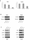
Anti-Wnt-1 antibody inhibited β-catenin/Tcf4 transcriptional activity. A). Tcf4 reporter assay of Tcf-dependent transcriptional activity in Huh7 and Hep40 cell lines. Huh7 and Hep40 cells were co-transfected with plasmid encoding β-galactosidase (a control for transfection efficiency) and either the pTOPFLASH or pFOPFLASH reporters. Cells were incubated with anti-Wnt-1 antibody or control IgG (2 μg/ml) and harvested after 48 hr to measure luciferase and β-galactosidase activities. Reporter gene activation is expressed in terms of relative light units (RLU) detected in pTOPFLASH or pFOPFLASH transfected cells and normalized for β-galactosidase activity. The results are expressed as mean ± SD (error bars). Experiments were performed in triplicates; P < 0.05. B). Anti-Wnt-1 antibody decreased nuclear β-catenin accumulation in Huh7 and Hep40 cells. Histone 3 was used as the loading control. C). The effect of anti-Wnt-1 antibody on the expression of β-catenin/Tcf4 target genes c-Myc, cyclin D1, and survivin. Huh7 and Hep40 cells were incubated for 48 hr with anti-Wnt-1 antibody (2 μg/ml) and c-Myc, cyclin D1, survivin and β-actin (loading control) levels were determined by Western blotting using specific antibodies.
Figure 4
Figure 4
Anti-Wnt-1 antibody suppressed tumor xenograft growth in vivo. A). Nude mice bearing Huh7 tumor xenografts were treated with anti-Wnt-1 antibody (dose: 50 μg/kg; once a week), or with PBS as a control (n = 5 in each group). Significant differences in the tumor volumes between anti-Wnt-1 antibody-treated and PBS control groups were observed at the beginning of the third week after initiation of treatment (P < 0.05). B). TUNEL staining of the xenograft specimens removed from controls and mice treated with anti-Wnt-1 antibody (200× magnification). Red arrows indicate the positively stained apoptotic cells. C). Xenograft specimens removed from controls and mice treated with anti-Wnt-1 antibody were immunostained to detect expression of c-Myc, cyclin D1, and survivin (400× magnification). The number of positively stained cells were counted from three randomly selected areas and the values represent mean ± SD (error bars).

References

    1. Parkin DM, Pisani P, Ferlay J. Estimates of the worldwide incidence of 25 major cancers in 1990. Int J Cancer. 1999;80:827–841. doi: 10.1002/(SICI)1097-0215(19990315)80:6<827::AID-IJC6>3.0.CO;2-P. - DOI - PubMed
    1. Llovet JM, Bustamante J, Castells A, Vilana R, Ayuso Mdel C, Sala M, Bru C, Rodes J, Bruix J. Natural history of untreated nonsurgical hepatocellular carcinoma: rationale for the design and evaluation of therapeutic trials. Hepatology. 1999;29:62–67. doi: 10.1002/hep.510290145. - DOI - PubMed
    1. Llovet JM, Burroughs A, Bruix J. Hepatocellular carcinoma. Lancet. 2003;362:1907–1917. doi: 10.1016/S0140-6736(03)14964-1. - DOI - PubMed
    1. Liu JH, Chen PW, Asch SM, Busuttil RW, Ko CY. Surgery for hepatocellular carcinoma: does it improve survival? Ann Surg Oncol. 2004;11:298–303. doi: 10.1245/ASO.2004.03.042. - DOI - PubMed
    1. Peifer M, Polakis P. Wnt signaling in oncogenesis and embryogenesis--a look outside the nucleus. Science. 2000;287:1606–1609. doi: 10.1126/science.287.5458.1606. - DOI - PubMed

Publication types

MeSH terms

LinkOut - more resources