Review
. 2010 May 1;341(1):34-55.
doi: 10.1016/j.ydbio.2009.09.024. Epub 2009 Sep 22.

Morphogenesis of epithelial tubes: Insights into tube formation, elongation, and elaboration

Affiliations
Review

Morphogenesis of epithelial tubes: Insights into tube formation, elongation, and elaboration

Deborah J Andrew et al. Dev Biol. .

Abstract

Epithelial tubes are a fundamental tissue across the metazoan phyla and provide an essential functional component of many of the major organs. Recent work in flies and mammals has begun to elucidate the cellular mechanisms driving the formation, elongation, and branching morphogenesis of epithelial tubes during development. Both forward and reverse genetic techniques have begun to identify critical molecular regulators for these processes and have revealed the conserved role of key pathways in regulating the growth and elaboration of tubular networks. In this review, we discuss the developmental programs driving the formation of branched epithelial networks, with specific emphasis on the trachea and salivary gland of Drosophila melanogaster and the mammalian lung, mammary gland, kidney, and salivary gland. We both highlight similarities in the development of these organs and attempt to identify tissue and organism specific strategies. Finally, we briefly consider how our understanding of the regulation of proliferation, apicobasal polarity, and epithelial motility during branching morphogenesis can be applied to understand the pathologic dysregulation of these same processes during metastatic cancer progression.


Figures

Figure 1
Figure 1
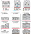
All epithelial tubes comprise polarized cells surrounding a central lumen, with the apical surfaces of the cells facing the lumen and the basal surface contacting either a basal lamina or other cells (A). Apical surfaces often contain microvilli, actin-rich, finger-like membranous structures that extend into the lumen. Epithelial cells contact each other through junctional structures that serve to link the cells, provide structural support, prevent the diffusion of membrane proteins and lipids, and provide barrier function. The arrangement of junctions varies slightly between mammalian and insect epithelia. Epithelia have been categorized based on the shapes of individual epithelial cells and the number and relative arrangement of cell layers (B). Some epithelial tubes comprise an inner epithelial layer and an outer “muscle” cell layer that is either directly contacting the epithelium or that is separated by a basal lamina (C).
Figure 2
Figure 2
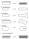
Epithelial tube formation involves dynamic cellular changes, beginning with epithelial polarization to form the cell layer that surrounds the central lumen (A). The geometry of cells in the tube primordium, coupled with the order and arrangement of cells undergoing morphogenesis will determine the shape of the initial polarized tube. Once the initial tube has formed, it will elongate using a variety of strategies (B). Variations in final form can be achieved during elongation by the processes of bifurcation (C), where the end of the tube splits into two smaller tubes, side branching (D), where a new tube will form on the side of the main tube branch, and clefting (E), where a division is created within the outgrowing tube primordia that either prevents outgrowth at a specific position or that actively splits cells into separate populations by the exchange of cell – cell junctions for cell – ECM junctions. Many tubes use a combination of mechanisms to achieve their final form. Final tube size is determined by the size of the primordium, cell growth, cell division and cell recruitment (F). In Drosophila, tube size regulation, both diameter and length, is linked to the secretion and modification of an apical matrix.
Figure 3
Figure 3
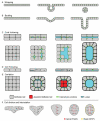
Tube formation can begin with an already polarized epithelium during the processes of wrapping (A) or budding (B). Tubes can also form from unpolarized rudiments that polarize and create a lumen by three proposed mechanisms. In cord hollowing, the cells polarize and create small lumenal spaces at sites of apical membrane contact. These spaces subsequently coalesce into a single common lumen (C). In cell hollowing, lumenal space is first created within individual cells by the formation of large apical vesicles. These vesicles eventually fuse with each other and with the plasma membrane to form a large common lumen (D). During cavitation, cells on the periphery of the primordium will form the epithelium and cells in the center will either die by apoptosis or intercalate into the periphery and contribute to the forming epithelium (E). The zebrafish neural tube forms by a mechanism of cell divisions that establish apico-basal polarity and cell intercalation to correctly position the apical and basal domains within the forming tube. The image in F was slightly modified from Tawk et al., 2007.
Figure 4
Figure 4
Tubes can elongate by several distinct mechanisms. Cell number can remain constant and tubes can elongate as individual cells become longer (A). Cell number can remain constant and tubes can elongate as cells rearrange (B). Cells can divide either locally (C) or globally (D) to increase tube length. New cells can be recruited to elongate the tube (E).
Figure 5
Figure 5
Tube elongation and elaboration can occur through several means. Tubes may elongate as a single chain of cells or as a group of cells that either remains fully or partially polarized. Tube elongation may involve single or multiple leaders and the tube may or may not be contained within a basal lamina. Cellular protrusions may occur on the basal surfaces of none, some or all of the epithelial cells.
Figure 6
Figure 6
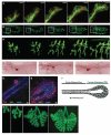
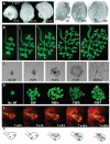
In vivo tube formation reveals a variety of strategies for tube elongation and elaboration. The Drosophila salivary gland invaginates through a budding mechanism (A; Live images provided by B. Kerman, A. Cheshire and D.J.A. (Cheshire et al., 2008). Expression of nuclear Ds-Red and α-catenin-GFP are driven by a fkh-Gal4 driver, which drives expression of UAS-constructs specifically in the salivary gland). The tube remains fully polarized as it elongates via cell shape change and migrates to its final position. The Drosophila trachea begins as polarized epithelia that internalize and form primary branches by a budding mechanism (B; Live images provided by B. Kerman, A. Cheshire and D.J.A. Expression of cytoplasmic GFP is driven by a btl-Gal4 driver, which drives expression of UAS-constructs throughout the trachea and in midline glial cells). The tracheae remain polarized during the process of branch migration and the tubes elongate by a combination of cell shape change and cell rearrangement. Mammalian tubes are often very complex structures and can elaborate through a variety of mechanisms. The mammary gland begins as an unpolarized rudiment that polarizes by cavitation (C; Fixed images were provided by A.J.E. and Z. Werb). The mammalian ducts are simple bilayered epithelia, with a polarized epithelial layer facing the lumen surrounded by a monolayer of myoepithelial cells that stain for smooth muscle actin (red) (D). Staining with phospho-histone H3 reveals limited cell divisions within the polarized epithelium (green). The terminal end buds are the units of elongation, which are an actively dividing relatively unpolarized group of cells, known to elongate by a mechanism known as collective cell migration (E; images provided by A. Ewald and reproduced with permission from Developmental Cell, Vol 14, pages 570-581). A cartoon diagram of the elongating mammary gland reveals the mature polarized duct and the complex, relatively unpolarized terminal end bud, which undergoes extensive proliferation (green cells). The mouse lung undergoes a stereotypical series of planar and orthogonal bifurcations and side branchings to achieve its final form. Unlike the mammary gland, the mammalian lung maintains its polarity during growth and branching morphogenesis. The pattern of mouse lung branching was determined by examining large numbers of developing lungs isolated at distinct developmental stages (G; images provided by R. Metzger and reproduced with permission from Nature Vol 453, pages 745-50).
Figure 7
Figure 7
As with the mammary gland, the mammalian salivary gland forms from an unpolarized rudiment that polarizes by cavitation. Clefting leads to the formation of the multiple buds of the fully formed glands. Salivary glands isolated directly from embryos on days E12, E13 and E14 show increased levels of branching with increased age (A). Similar patterns of branching are observed with salivary glands cultured in vivo (salivary gland images provided by M. Hoffman, and reproduced with permission from Differentiation Vol 74, Pages 349-364). The mouse ureteric buds can be cultured ex vivo and through the use of tissue-specific GFP markers imaged live to follow the process of tube elaboration (B; images provided by O. Michos and F. Costantini). The ureteric bud derives from the posterior portion of the Wolffian duct and undergoes a series of bifurcations, trifurcations and side branchings to give rise to the elaborate architecture of the mammalian kidney. Explant cultures of salivary glands (C; images provided by M. Hoffman; FGF1 was at 100 ng/ml, FGF2 and FGF7 at 200 ng/ml, and FGF10 at 500 ng/ml) and mammary glands (D; images provided by A.J.E. and Z. Werb; 2.5 nM of indicated growth factors) in which the mesenchyme has been removed are excellent substrates for imaging tube formation and for directly testing of the effects of different growth factors and inhibitors on tube architecture. Organotypic cultures of mammary glands containing both the epithelial cells and surrounding myoepithelial cells can be used to determine the roles of cell interactions on organ morphogenesis (E; images provided by A.J.E. and Z. Werb). Individual cells are outlined and followed during tube elongation (E’).

Similar articles

Cited by

References

    1. Abrams EW, Mihoulides WK, Andrew DJ. Fork head and Sage maintain a uniform and patent salivary gland lumen through regulation of two downstream target genes, PH4alphaSG1 and PH4alphaSG2. Development. 2006;133:3517–27. - PubMed
    1. Adler PN. Planar signaling and morphogenesis in Drosophila. Dev Cell. 2002;2:525–35. - PubMed
    1. Ainsworth C, Wan S, Skaer H. Coordinating cell fate and morphogenesis in Drosophila renal tubules. Philos Trans R Soc Lond B Biol Sci. 2000;355:931–7. - PMC - PubMed
    1. Araujo SJ, Aslam H, Tear G, Casanova J. mummy/cystic encodes an enzyme required for chitin and glycan synthesis, involved in trachea, embryonic cuticle and CNS development--analysis of its role in Drosophila tracheal morphogenesis. Dev Biol. 2005;288:179–93. - PubMed
    1. Bagnat M, Cheung ID, Mostov KE, Stainier DY. Genetic control of single lumen formation in the zebrafish gut. Nat Cell Biol. 2007;9:954–60. - PubMed

LinkOut - more resources