

MBT domain proteins in development and disease
- PMID: 19778625
- PMCID: PMC3772645
- DOI: 10.1016/j.semcdb.2009.09.010
MBT domain proteins in development and disease
Abstract
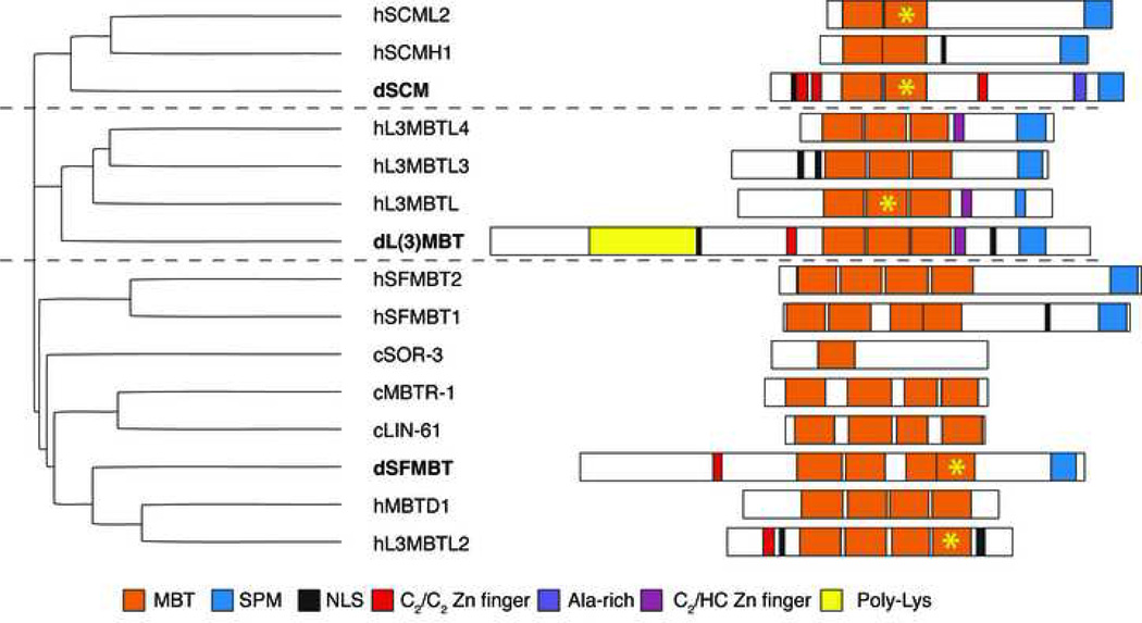
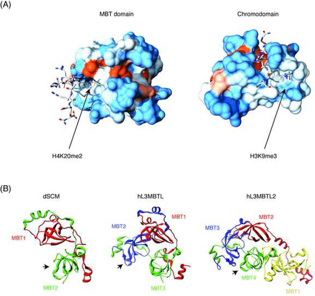
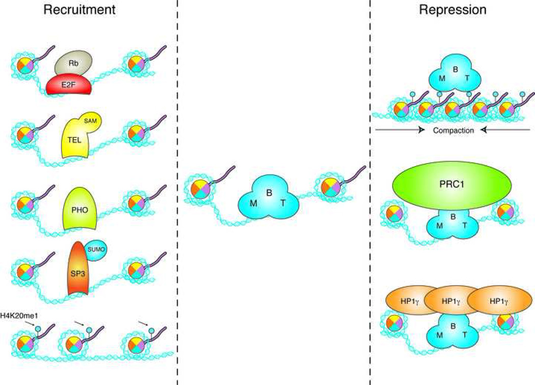
The Malignant Brain Tumor (MBT) domain is a "chromatin reader", a protein module that binds to post-translational modifications on histone tails that are thought to affect a variety of chromatin processes, including transcription. More specifically, MBT domains recognize mono- and di-methylated lysines at a number of different positions on histone H3 and H4 tails. Three Drosophila proteins, SCM, L(3)MBT and SFMBT contain multiple adjacent MBT repeats and have critical roles in development, maintenance of cell identity, and tumor suppression. Although they function in different pathways, these proteins all localize to chromatin in vivo and repress transcription by a currently unknown molecular mechanism that requires the MBT domains. The human genome contains several homologues of these MBT proteins, some of which have been linked to important gene regulatory pathways, such as E2F/Rb- and Polycomb-mediated repression, and to the insurgence of certain neurological tumors. Here, we review the genetics, biochemistry, and cell biology of MBT proteins and their role in development and disease.
Copyright 2009 Elsevier Ltd. All rights reserved.
Figures
References
-
- Kouzarides T. Chromatin Modifications and Their Function. Cell. 2007;128:693–705. - PubMed
-
- Jenuwein T, Allis CD. Translating the histone code. Science. 2001;293:1074–1080. - PubMed
-
- Ruthenburg AJ, Allis CD, Wysocka J. Methylation of lysine 4 on histone H3: intricacy of writing and reading a single epigenetic mark. Mol Cell. 2007;25:15–30. - PubMed
-
- Sims RJ, Reinberg D. Histone H3 Lys 4 methylation: caught in a bind? Genes Dev. 2006;20:2779–2786. - PubMed
Publication types
MeSH terms
Substances
Grants and funding
LinkOut - more resources
Full Text Sources
Other Literature Sources
Medical
Molecular Biology Databases
