. 2009 Dec 25;284(52):36312-36323.
doi: 10.1074/jbc.M109.049817. Epub 2009 Sep 24.

DGAT1 expression increases heart triglyceride content but ameliorates lipotoxicity

Affiliations

DGAT1 expression increases heart triglyceride content but ameliorates lipotoxicity

Li Liu et al. J Biol Chem. .

Abstract

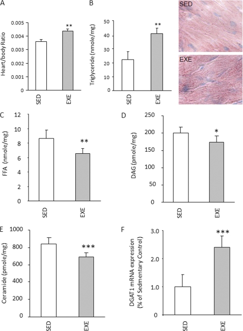
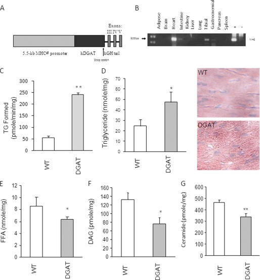
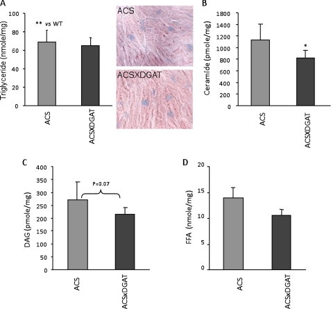
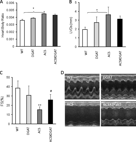
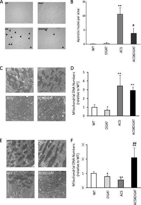
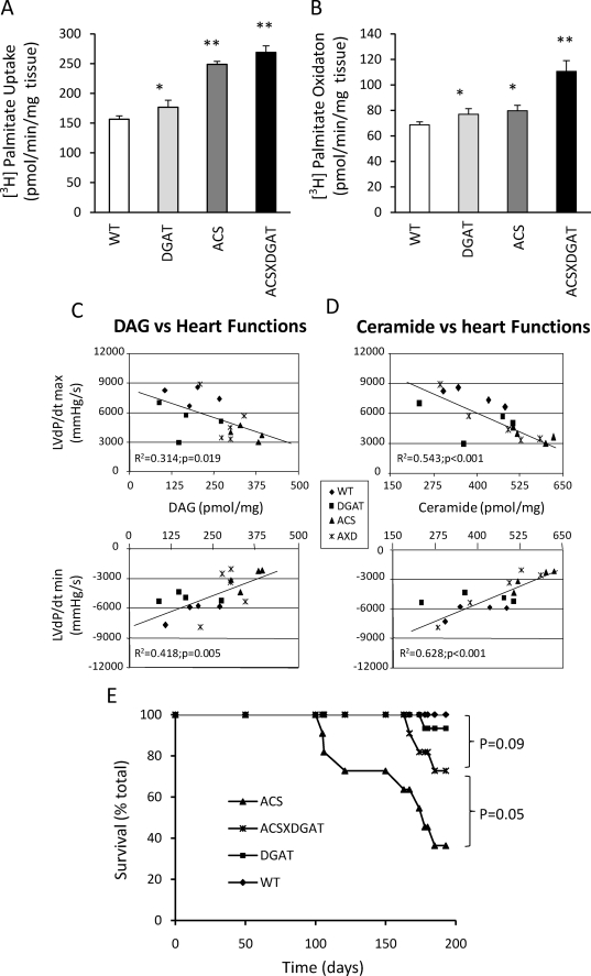
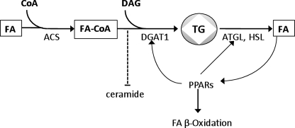
Intracellular lipid accumulation in the heart is associated with cardiomyopathy, yet the precise role of triglyceride (TG) remains unclear. With exercise, wild type hearts develop physiologic hypertrophy. This was associated with greater TG stores and a marked induction of the TG-synthesizing enzyme diacylglycerol (DAG) acyltransferase 1 (DGAT1). Transgenic overexpression of DGAT1 in the heart using the cardiomyocyte- specific alpha-myosin heavy chain (MHC) promoter led to approximately a doubling of DGAT activity and TG content and reductions of approximately 35% in cardiac ceramide, 26% in DAG, and 20% in free fatty acid levels. Cardiac function assessed by echocardiography and cardiac catheterization was unaffected. These mice were then crossed with animals expressing long-chain acyl-CoA synthetase via the MHC promoter (MHC-ACS), which develop lipotoxic cardiomyopathy. MHC-DGAT1XMHC-ACS double transgenic male mice had improved heart function; fractional shortening increased by 74%, and diastolic function improved compared with MHC-ACS mice. The improvement of heart function correlated with a reduction in cardiac DAG and ceramide and reduced cardiomyocyte apoptosis but increased fatty acid oxidation. In addition, the survival of the mice was improved. Our study indicates that TG is not likely to be a toxic lipid species directly, but rather it is a feature of physiologic hypertrophy and may serve a cytoprotective role in lipid overload states. Moreover, induction of DGAT1 could be beneficial in the setting of excess heart accumulation of toxic lipids.


Figures

FIGURE 1.
FIGURE 1.
Exercise effects on heart DGAT1 expression and lipid. A, heart to body weight ratio in sedentary (SED) and exercised (EXE) FVB mice (n = 5). B–E, TG, FFA, DAG, and ceramide content in heart muscles isolated from sedentary and exercised FVB mice (n = 5). F, relative DGAT1 expression in exercise group compared with sedentary group, real time PCR (n = 5). *, p < 0.05; **, p < 0.01; ***, p < 0.001.
FIGURE 2.
FIGURE 2.
Creation and characterization of MHC-DGAT1 transgenic mice. A, DGAT1 transgene contains (from the 5′- to 3′-end) the 5.5-kb MHC promoter, a human DGAT1 cDNA containing its own initiation and termination codons, and the genomic sequence of human growth hormone (hGH) containing the last 3 exons and 2 introns as indicated. B, tissue distribution of the DGAT1 transgene mRNA levels measured by reverse transcription and PCR amplification. C, total DGAT activity levels in membrane fractions of heart muscles from WT and transgenic mice (n = 3 in each group). D–G, TG, FFA, DAG, and ceramide contents in heart muscles isolated from WT and transgenic mice (n = 5–6). *, p < 0.05; **, p < 0.01.
FIGURE 3.
FIGURE 3.
Comparison of cardiac lipids in MHC-ACS and MHC-ACSXMHC-DGAT1 mice. A–D, TG, FFA, DAG, and ceramide concentrations in heart muscles isolated from MHC-ACS and MHC-ACSXMHC-DGAT1 transgenic mice (n = 8–11).
FIGURE 4.
FIGURE 4.
Cardiac function in MHC-DGAT1 and MHC-DGAT1/MHC-ACS mice. A, heart to body weight ratio in WT, MHC-DGAT1, MHC-ACS, and MHC-DGAT1/MHC-ACS transgenic mice (n = 5–7). Echocardiography showed left ventricular systolic dimension (B) and fractional shortening (C). Photographs are shown of echocardiograms (D) (n = 5–7). **, p < 0.01 MHC-ACS versus WT or MCH-DGAT1; #, p < 0.05 MHC-ACS versus MHC-ACS/MHC-DGAT1.
FIGURE 5.
FIGURE 5.
Apoptosis and mitochondrial analysis. A, histologic examination of the hearts by terminal dUTP nick-end labeling staining for WT, MHC-DGAT1, MHC-ACS, and MHC-DGAT1X MHC-ACS transgenic mice (A) and quantification of the apoptotic cells (B) (n = 3). Data are shown as mean ± S.D. *, p < 0.05; **, p < 0.01; #, p < 0.05; ##, p < 0.01 versus MHC-ACS. Arrows indicate apoptotic cells. C, electron micrograph; D, quantification of mitochondria in 4-month-old mice. E, electron micrograph; F, and quantification of mitochondria in 6-month-old mice.
FIGURE 6.
FIGURE 6.
Fatty acid uptake and oxidation, correlation of lipids and heart function, and survival rates. A and B, wild type and transgenic mice (MHC-DGAT1, MHC-ACS, and MHC-DGAT1XMHC-ACS) (n = 4) heart slices were incubated with [3H]palmitate acid as described under “Experimental Procedures.” C, positive correlation between heart muscle DAG levels with left ventricle contractile function (mm Hg/s), (adjusted R2 = 0.314; p = 0.019) and with diastolic function (mm Hg/s) (adjusted R2 = 0.418; p = 0.005). D, positive correlation between heart muscle ceramide levels with left ventricle contractile function (mm Hg/s) (adjusted R2 = 0.543; p < 0.001) and with diastolic function (mm Hg/s) (adjusted R2 = 0.628; p ≤ 0.001). E, wild type (WT, n = 10) and transgenic mice (WT, n = 10; MHC-DGAT, n = 15; MHC-ACS, n = 11; and MHC-ACSXMHC-DGAT1, n = 11) were followed for 180 days, and incidence of spontaneous death was recorded as a function of time. *, p < 0.5; **, p < 0.01.
FIGURE 7.
FIGURE 7.
DGAT1, ATGL, and PPAR regulation. Increased TG droplet storage induced by DGAT1 is associated with greater ATGL expression. Subsequent hydrolysis of TG provides agonists that lead to greater expression of PPAR downstream genes and greater fatty acid oxidation.

References

    1. Cases S., Smith S. J., Zheng Y. W., Myers H. M., Lear S. R., Sande E., Novak S., Collins C., Welch C. B., Lusis A. J., Erickson S. K., Farese R. V., Jr. (1998) Proc. Natl. Acad. Sci. U.S.A. 95, 13018–13023 - PMC - PubMed
    1. Yen C. L., Monetti M., Burri B. J., Farese R. V., Jr. (2005) J. Lipid Res. 46, 1502–1511 - PubMed
    1. Oelkers P., Behari A., Cromley D., Billheimer J. T., Sturley S. L. (1998) J. Biol. Chem. 273, 26765–26771 - PubMed
    1. Lardizabal K. D., Mai J. T., Wagner N. W., Wyrick A., Voelker T., Hawkins D. J. (2001) J. Biol. Chem. 276, 38862–38869 - PubMed
    1. Meegalla R. L., Billheimer J. T., Cheng D. (2002) Biochem. Biophys. Res. Commun. 298, 317–323 - PubMed

Publication types

MeSH terms