. 2009 Nov;37(21):7047-58.
doi: 10.1093/nar/gkp765.

Two Dot1 isoforms in Saccharomyces cerevisiae as a result of leaky scanning by the ribosome

Affiliations

Two Dot1 isoforms in Saccharomyces cerevisiae as a result of leaky scanning by the ribosome

Floor Frederiks et al. Nucleic Acids Res. 2009 Nov.

Abstract

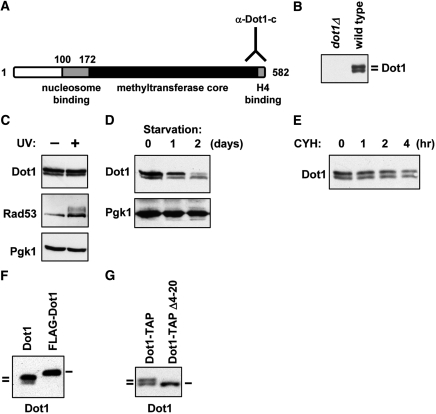
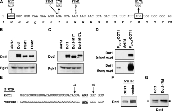
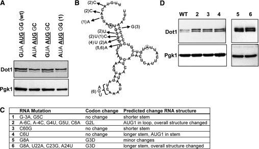
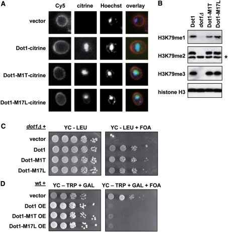
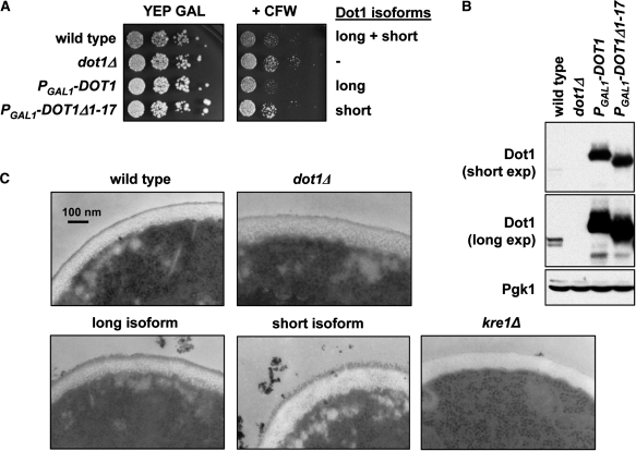
Dot1 is a conserved histone methyltransferase that methylates histone H3 on lysine 79. We previously observed that in Saccharomyces cerevisiae, a single DOT1 gene encodes two Dot1 protein species. Here, we show that the relative abundance of the two isoforms changed under nutrient-limiting conditions. A mutagenesis approach showed that the two Dot1 isoforms are produced from two alternative translation start sites as a result of leaky scanning by the ribosome. The leaky scanning was not affected by the 5'- or 3'-untranslated regions of DOT1, indicating that translation initiation is determined by the DOT1 coding sequence. Construction of yeast strains expressing either one of the isoforms showed that both were sufficient for Dot1's role in global H3K79 methylation and telomeric gene silencing. However, the absence of the long isoform of Dot1 altered the resistance of yeast cells to the chitin-binding drug Calcofluor White, suggesting that the two Dot1 isoforms have a differential function in cell wall biogenesis.


Figures

Figure 1.
Figure 1.
Formation of two Dot1 isoforms depends on the N-terminus. (A) Schematic overview of the structure of the yeast Dot1 protein with a lysine-rich putative nucleosome binding domain, a methyltransferase core and a C-terminal domain that binds to histone H4. The Dot1 antibody is directed against the very C-terminus of the protein. Numbering of amino acid residues starts at the first methionine, a residue that is most likely absent from the mature protein due to co-translational cleavage. (B) Immunoblot analysis of Dot1 expression shows that two isoforms are present in log phase cultures. Whole-cell extracts of a dot1Δ strain (UCC7183) transformed with an empty plasmid (pRS315) or a single copy plasmid expressing DOT1 (pDOT1) were analyzed. (C) Immunoblot analysis of Dot1 expression in wild-type cells (NKI3031) following UV irradiation of cells arrested in G1. The mobility shift of Rad53 caused by UV-induced phosphorylation was used to confirm activation of the DNA-damage checkpoint. Pgk1 was used as a loading control. (D) Whole-cell lysates of wild-type strain NKI3031 were prepared from exponentially growing cultures and cultures of starved cells grown overnight or over two nights. Dot1 expression was analyzed by immunoblot and Pgk1 was used as a loading control. (E) To analyze the protein half life of the two Dot1 isoforms, 50 µg/ml cycloheximide was added to a culture of exponentially growing dot1Δ cells transformed with a DOT1 overexpression plasmid (UCC7183 + pFvL18). At the indicated time points aliquots of the culture were taken for immunoblot analysis of Dot1 expression. (F) Immunoblot analysis of Dot1 expression in cells expressing full-length Dot1 without or with an N-terminal FLAG epitope tag (UCC7183 + pDOT1 or pFvL201). (G) Dot1 expression in cells containing full-length Dot1 (UCC7183 + pFvL212) or a deletion mutant of Dot1 (Δ4-20, UCC7183 + pFvL215) was analyzed by immunoblot. Both proteins have a C-terminal TAP tag.
Figure 2.
Figure 2.
The two Dot1 isoforms are generated by leaky scanning by the ribosome, which is not dependent on the 5′-context of the start codon. (A) Nucleotide and amino acid sequence of the N-terminal part of Dot1. Black boxes show the two potential start codons. The different mutants used in this study are shown underlined. Briefly, in the M1T mutant AUG1 is changed to ACU, in the I7M mutant AUA7 is changed to AUG and in the M17L mutant AUG17 is changed to UUG. FSM1 has an insertion of an A in codon 9 and FSM2 has an insertion of a C in codon 7. Sequence details of the FSMs are shown in Table 1. (B) Immunoblot analysis of FSMs of Dot in a dot1Δ strain (UCC7183) transformed with an empty plasmid (pRS315), a single copy plasmid expressing DOT1 (pDOT1), or a single copy plasmid expressing one of the two FSMs of DOT1 (pGH18 or pGH21). Pgk1 was used as a loading control. (C) Immunoblot analysis of Dot1 expression in dot1Δ cells (UCC7183) containing an empty plasmid, wild-type Dot1, Dot1-M1T or Dot1-M17L (pRS315, pDOT1, pGH01 and pGH02, respectively), showing that mutation of either of the putative start codons leads to expression of a single Dot1 isoform. Pgk1 was used as a loading control. (D) Comparison of Dot1 expression in a wild-type strain (PDOT1-DOT1, UCC7164) and a dot1Δ strain (UCC7183) containing an empty vector (pTCG) or a plasmid overexpressing DOT1 (and 54 bp of its 5′-UTR) from the GAL1 promoter (PGAL1-DOT1, pFvL18). A short and a long exposure of the same blot are shown. (E) Nucleotide sequence around the DOT1 start codon in strains NKI3049 (endogenous DOT1 5′-UTR) and NKI3066 (non-yeast vector 5′-UTR). Thirty nucleotides of 5′-UTR sequence are shown. In strain NKI3049 (DOT1) 62 bp of endogenous DOT1 5′-UTR remains upstream of the first start codon; in strain NKI3066 (vector) the DOT1 5′-UTR has been removed by insertion of the GAL1 promoter and 45 bp of vector sequence of the integration construct pYM-N22. (F) Immunoblot comparison of Dot1 expression in the two strains described in E. (G) Immunoblot analysis of Dot1 expression in a dot1Δ strain (UCC7183) transformed with wild-type Dot1 (pDOT1) or Dot1-I7M (pFF018).
Figure 3.
Figure 3.
Effect of sequences proximal to the start codon on leaky scanning. (A) Immunoblot analysis of Dot1 expression in strains in which the context of AUG1 has been mutated. The nucleotide sequence is indicated above the lanes. Wild-type cells (UCC7164) are compared with cells harboring the mutation G–3→A–3, G + 5→C + 5, or the double mutation (UCC7183 + pFvL025, pFvL026 and pFvL027, respectively). Numbers are relative to the first nucleotide of AUG1. Pgk1 was used as a loading control. (B) RNA structure of DOT1 mRNA (–10 to +65) predicted by RNAfold (27). Solid circle indicates start of AUG1, dashed circle indicates start of AUG17. Mutant numbers refer to panel C (2–6). (C) Summary of mutations in DOT1 mRNA. (D) Dot1 expression of mutants shown in panel C. Numbers 1–4 are mutants of single copy pDOT1, number 6 is a mutant of PGAL1-DOT1 (number 5) which already contained a G8A mutation.
Figure 4.
Figure 4.
The N-terminus of Dot1 does not affect localization, H3K79 methylation or telomeric silencing. (A) Analysis of the localization of wild-type Dot1, Dot1-M1T and Dot1-M17L. Log-phase cells overexpressing one of these proteins fused to a fluorescent citrine tag were stained with ConA-Cy5 (cell wall) and Hoechst (DNA) and then imaged using a confocal microscope. For this experiment dot1Δ strain UCC7183 was transformed with pFvL221, pTW055, pGH024 and pGH025. (B) Whole-cell extracts from log-phase cultures were prepared from dot1Δ strain UCC7183 transformed with an empty plasmid or a single copy plasmid expressing Dot1, Dot1-M1T or Dot1-M17L (pRS315, pDOT1, pGH01 and pGH02, respectively). H3K79 methylation and total histone H3 were analyzed by immunoblot. The asterisk indicates a non-specific band recognized by the H3K79me2 antibody. (C) Telomeric silencing of a URA3 reporter gene close to telomere VIIL was measured in the strains described in (B). Cells were plated in 10-fold dilution series on selective media in the presence or absence of 1 g/l 5-FOA. Cells that silence URA3 are resistant to FOA; cells with loss of telomeric silencing express URA3 and are sensitive to FOA. (D) Analysis of telomeric silencing in a wild-type strain (UCC7164) transformed with pTCG, pTCG-DOT1 (pFvL18), pTCG-dot1-M1T (pGH03) or pTCG-dot1-M17L (pGH04). The GAL1 promoter on the pTCG plasmid was induced by growth on selective media containing galactose (GAL) to overexpress (OE) the Dot1 proteins.
Figure 5.
Figure 5.
Dot1 is involved in cell wall function. (A) Sensitivity to CFW of the following strains: wild type (UCC7164), dot1Δ (UCC7183), PGAL1-3xHA-DOT1 (NKI1059) and PGAL1-3xHA-DOT1Δ1-17 (NKI1060). Cells were plated in 10-fold dilutions on rich media containing 2% galactose in the presence or absence of 20 µg/ml CFW. The image is representative of four independent experiments. (B) Immunoblot analysis of Dot1 expression in the strains described in (A). A long and short exposure is shown to indicate the expression relative to endogenous Dot1. Pgk1 was used as a loading control. (C) The cell wall of the strains described in (A) was imaged using electron microscopy. A kre1Δ strain (YSC-1122) was used as a control.

Similar articles

Cited by

References

    1. van Leeuwen F, Gafken PR, Gottschling DE. Dot1p modulates silencing in yeast by methylation of the nucleosome core. Cell. 2002;109:745–756. - PubMed
    1. Ng HH, Feng Q, Wang H, Erdjument-Bromage H, Tempst P, Zhang Y, Struhl K. Lysine methylation within the globular domain of histone H3 by Dot1 is important for telomeric silencing and Sir protein association. Genes Dev. 2002;16:1518–1527. - PMC - PubMed
    1. Lacoste N, Utley RT, Hunter JM, Poirier GG, Cote J. Disruptor of telomeric silencing-1 is a chromatin-specific histone H3 methyltransferase. J. Biol. Chem. 2002;277:30421–30424. - PubMed
    1. Feng Q, Wang H, Ng HH, Erdjument-Bromage H, Tempst P, Struhl K, Zhang Y. Methylation of H3-lysine 79 is mediated by a new family of HMTases without a SET domain. Curr. Biol. 2002;12:1052–1058. - PubMed
    1. Wysocki R, Javaheri A, Allard S, Sha F, Cote J, Kron SJ. Role of Dot1-dependent histone H3 methylation in G1 and S phase DNA damage checkpoint functions of Rad9. Mol. Cell Biol. 2005;25:8430–8443. - PMC - PubMed

Publication types

MeSH terms

Substances