. 2009 Nov 13;326(5955):1005-7.
doi: 10.1126/science.1180962. Epub 2009 Sep 24.

Amyloid-beta dynamics are regulated by orexin and the sleep-wake cycle

Affiliations

Amyloid-beta dynamics are regulated by orexin and the sleep-wake cycle

Jae-Eun Kang et al. Science. .

Abstract

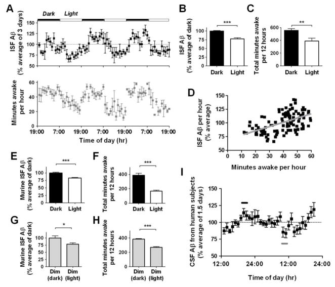
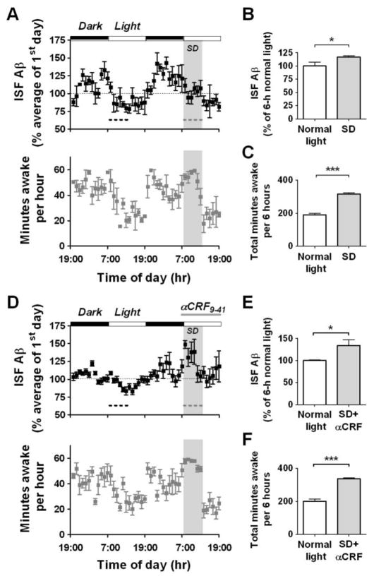
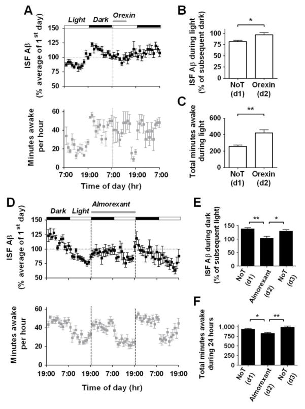
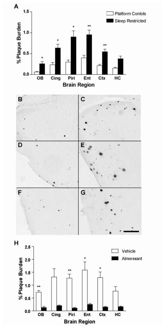
Amyloid-beta (Abeta) accumulation in the brain extracellular space is a hallmark of Alzheimer's disease. The factors regulating this process are only partly understood. Abeta aggregation is a concentration-dependent process that is likely responsive to changes in brain interstitial fluid (ISF) levels of Abeta. Using in vivo microdialysis in mice, we found that the amount of ISF Abeta correlated with wakefulness. The amount of ISF Abeta also significantly increased during acute sleep deprivation and during orexin infusion, but decreased with infusion of a dual orexin receptor antagonist. Chronic sleep restriction significantly increased, and a dual orexin receptor antagonist decreased, Abeta plaque formation in amyloid precursor protein transgenic mice. Thus, the sleep-wake cycle and orexin may play a role in the pathogenesis of Alzheimer's disease.


Figures

Figure 1
Figure 1
Diurnal rhythm of ISF Aβ levels in the hippocampus of mice and CSF Aβ levels in human subjects. (A) ISF human Aβ levels expressed as a percentage of basal ISF Aβ levels over 6 light-dark periods in Tg2576 mice (n = 8). Total number of minutes spent awake per hour in the same mice. (B, C) Mean ISF Aβ levels were 24.4% higher (***P < 0.0001, n = 8) and the number of minutes awake were 167 minutes higher (**P = 0.007, n = 7) during dark vs. light periods. (D) ISF Aβ levels correlate with the number of minutes awake per hour (r = 0.53, ***P < 0.0001, n = 7). (E, F) Mean ISF murine Aβ levels and minutes awake over 2 days in C57BL6 mice. Under 12 hr dark/12 hr light conditions, ISF murine Aβ levels were 18.5% higher (***P < 0.0001, n = 10) and the number of minutes awake was 223 minutes higher (***P =0.0001, n = 5) during the dark periods. (G, H) Under constant dim light conditions, ISF Aβ levels were 22.7% higher (*P = 0.05, n = 10) and the number of minutes awake was 114 minutes higher (***P =0.0003, n = 5) during the typical hours for the dark phase. (I) CSF Aβ1–40 levels from human subjects expressed as a percentage of basal CSF Aβ1–40 levels over 33 hours (n = 10). Mean peak CSF Aβ1–40 levels (black bar) at 8–10 PM were 27.6% higher than mean trough CSF Aβ levels (gray bar) at 9–10 AM (112.3 ± 6% vs. 84.7 ± 6% respectively, P = 0.004). Data shown are mean ± SEM.
Figure 2
Figure 2
Acute sleep deprivation alters ISF Aβ diurnal rhythm independently of CRF receptor signaling in Tg2576 mice. (A) Mice underwent acute sleep deprivation (SD – grey dashed line) for 6 hours at the beginning of the light period. This prevented the normal decrease in ISF Aβ levels that occurs during this period (n = 8). (B) Mean ISF Aβ levels during SD were 16.8% higher compared to those during the light period 24 hours earlier (black dashed line, P = 0.05, n = 8). (C) Animals spent 126 more minutes awake during SD (***P < 0.0001, n = 5). (D) Mice underwent acute SD (grey dashed line) at the beginning of the light period. 860 pmoles of αCRF9–41 was infused into the hippocampus from 30 min before SD until the end of the light period (n = 8). (E) Mean ISF Aβ levels during SD with αCRF9–41 infusion were 33.7% higher compared to those during the light period 24 hours earlier (black dashed line, P = 0.01, n = 8). (F) Mice spent 136 more minutes awake during SD with αCRF9–41 infusion (***P < 0.0001, n = 5). Data represent mean ± SEM. SD = sleep deprivation.
Figure 3
Figure 3
Effects of orexin and a dual orexin receptor antagonist on ISF Aβ levels in Tg2576 mice. (A) After 24 hr of baseline measurement, 1.5 pmole/hr of orexin was infused icv for 6 hours at the beginning of the light period. This sustained ISF Aβ levels from the dark period, and kept the mice awake longer. (B, C) Infusion with orexin increased Aβ levels duirng the light period and thereby abolished the normal 20% difference between the dark and light period (*P = 0.01, n = 7). Orexin increased the amount of minutes awake by 163 minutes (**P = 0.009, n = 5), compared to that during the light period 24 hours previously. (D) After 24 hr of baseline measurement, 13.9 nmole/hr of almorexant was infused icv for 24 hr from the beginning of the dark period (n = 8). This continued to suppress ISF Aβ levels from the light period. (E) Mean ISF Aβ levels differed by 29% between the dark and light period during the control days, whereas there was no difference between the dark and light period during the 24 hour infusion of almorexant (**P = 0.001, n = 8). (F) During almorexant treatment, the number of minutes spent awake was decreased by 108 minutes over the 24 hour period compared to control days (**P = 0.005, n = 13). Data represent mean ± SEM. NoT = no treatment; d = day.
Figure 4
Figure 4
Aβ plaque deposition after chronic sleep restriction and chronic orexin receptor blockade in APPswe/PS1dE9 transgenic mice (A) Mice that underwent chronic sleep restriction for 21 days showed significantly greater Aβ plaque deposition in multiple subregions of the cortex compared to age-matched control mice (**P < 0.0008, *P < 0.008, n = 9–11 per group, using Bonferroni-adjusted P<0.0083 for multiple t-tests. For hippocampus, P<0.009). Representative photomicrographs of Aβ plaques are shown in (B) control and (C) sleep restricted olfactory bulb (D) control and (E) sleep restricted piriform cortex, (F) control and (G) sleep restricted entorhinal cortex. (H) Mice treated with daily i.p. injections of almorexant for 8 weeks showed significantly less Aβ plaque deposition in multiple subregions of the cortex compared to age-matched vehicle controls (**P < 0.0008, *P < 0.008 n = 5 per group, using Bonferroni-adjusted P<0.0083 for multiple t-tests. For cingulate cortex and hippocampus, P<0.009). Scale bar = 200 μm. OB = olfactory bulb, Cing = cingulate cortex, Piri = piriform cortex, Ent = entorhinal cortex, Ctx = cortex (immediately dorsal to dorsal hippocampus), and HC = hippocampus.

References

    1. Selkoe DJ. Nat Cell Biol. 2004 Nov;6:1054. - PubMed
    1. Kamenetz F, et al. Neuron. 2003 Mar 27;37:925. - PubMed
    1. Cirrito JR, et al. Neuron. 2005 Dec 22;48:913. - PubMed
    1. Hsiao K, et al. Science. 1996;274:99. - PubMed
    1. Kang JE, Cirrito JR, Dong H, Csernansky JG, Holtzman DM. Proc Natl Acad Sci U S A. 2007 Jun 19;104:10673. - PMC - PubMed

Publication types

MeSH terms