. 2009 Nov 18;28(22):3523-33.
doi: 10.1038/emboj.2009.283. Epub 2009 Sep 24.

RNase Y, a novel endoribonuclease, initiates riboswitch turnover in Bacillus subtilis

Affiliations

RNase Y, a novel endoribonuclease, initiates riboswitch turnover in Bacillus subtilis

Karen Shahbabian et al. EMBO J. .

Abstract

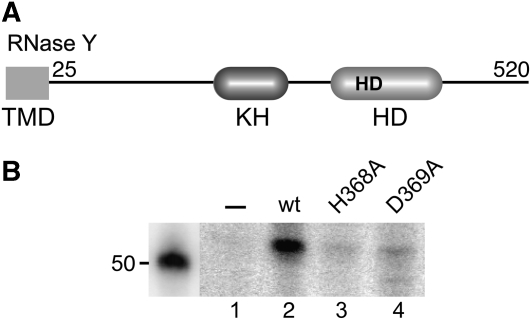
In contrast to Escherichia coli, initiation of mRNA decay in Gram-positive organisms is poorly understood. We studied the fate of the highly structured RNAs generated by premature transcription termination of S-adenosylmethionine (SAM)-dependent riboswitches in Bacillus subtilis. An essential protein of earlier unknown function, YmdA, was identified as a novel endoribonuclease (now called RNase Y) that was capable of preferential cleaving in vitro of the 5' monophosphorylated yitJ riboswitch upstream of the SAM-binding aptamer domain. Antiterminated full-length yitJ mRNA was not a substrate for RNase Y in vivo and in vitro, transcripts capable of forming the antiterminator were only cleaved in the presence of SAM. Turnover of 10 other SAM-dependent riboswitches was also initiated by RNase Y. Depletion of this ribonuclease increased the half-life of bulk mRNA more than two-fold. This indicates that RNase Y might be not only important for riboswitch RNA turnover but also as a key player in the initiation of mRNA decay in B. subtilis. About 40% of the sequenced eubacterial species have an RNase Y orthologue.


Conflict of interest statement

The authors declare that they have no conflict of interest.

Figures

Figure 1
Figure 1
Model for regulation of S-box gene expression in response to SAM. (A) In the presence of methionine (high SAM level), binding of SAM (represented by an encircled S) stabilizes the antiantiterminator structure favouring the formation of the terminator structure, which causes premature termination of transcription. (B) When SAM levels are low, the default antiterminator structure forms by base pairing of sequences of the antiantiterminator (grey, named I) and the terminator (hatched, named II), allowing expression of the downstream coding region. The positions of oligonucleotides are shown. Relevant positions on the transcript are numbered from the transcription start point (determined in Figure 2C). The yitJ AUG start codon is indicated.
Figure 2
Figure 2
Analysis of yitJ leader RNA in vivo. (A) Northern blot analysis of yitJ mRNA. Total RNA from the methionine auxotrophic strain SSB441, isolated from cultures grown in the presence or absence of methionine were separated on a denaturing 1.2% agarose gel. The RNA was probed with a uniformly labelled antisense 195 nt RNA covering 3′ sequences of the leader transcript and extending into the yitJ coding region (positions 152–346 Figure 1B delimited by oligonucleotides HP1231 and HP1056). An RNA sequence ladder was run in parallel as a size marker. Migration of 23S and 16S ribosomal RNAs is indicated. (B) High resolution Northern analysis of the yitJ leader. Total RNA from an RNase J1/J2 double mutant (rnjA inducible), an rnc mutant and a ymdA mutant (inducible), was isolated in the presence or absence of inducer, separated on an 8% polyacrylamide gel and probed with the leader-specific oligonucleotide HP1400 (Figure 1A). A [32P]-labelled DNA ladder was transferred to the membrane together with the RNA. (C) Identification of the yitJ transcription start. Oligonucleotide HP1238 (Figure 1A) was hybridized to and extended on total RNA isolated from a wild type and Pspac-ymdA mutant strain (±IPTG). The same primer was used for generating the sequence ladder. +1 indicates the band corresponding to the yitJ transcription start; the first transcribed nucleotide of the yitJ mRNA is encircled. (D) Primer extension analysis of total RNA isolated from a methionine auxotrophic strain grown in the presence or absence of methionine. T indicates the region corresponding to the terminator stem. (E) Sequence of the yitJ sigma A type promoter preceding the new transcription start point identified in (C).
Figure 3
Figure 3
Endonucleolytic cleavage of yitJ leader transcripts by YmdA. (A) Cleavage of a 5′ end-labelled monophosphorylated transcript (3′ end defined by primer HP1238). The major cleavage site was mapped on a sequence ladder generated with a primer whose 5′ end corresponded to the transcription start point. (B) Effect of increasing SAM concentrations (25 and 80 μM) on the cleavage of a 5′ end-labelled monophosphorylated transcript (3′ end defined by primer HP1356). (C) Cartoon of the riboswitch leader. The vertical arrow indicates the cleavage site for YmdA in the leader mRNA as identified in panel A. The position of the primers delimiting the transcript 3′ ends on the PCR templates are shown. The encircled S stands for SAM.
Figure 4
Figure 4
RNase Y cleavage of the yitJ riboswitch is sensitive to the 5′ phosphorylation state of the substrate. (A) Equal amounts of [32P] uniformly labelled tri- and monophosphorylated yitJ transcripts (3′ end defined by primer HP1238) were used as substrates in post-transcriptional cleavage assays with RNase Y as described in Material and methods. Reaction products were separated on an 8% polyacrylamide gel. The major cleavage site is indicated by scissors and the cleavage products are identified by schematic drawings on the right. A [32P]-labelled 50 bp DNA ladder was used as a size marker. (B) Kinetic analysis of RNase Y cleavage of 3′ end-labelled tri- and monophosphorylated yitJ transcripts (3′ end defined by primer HP1238). Samples from scaled-up cleavage assays were taken at the indicated time points. The reaction products were separated on a 5% polyacrylamide gel. The RNase Y-dependent cleavage product of 150 bases is indicated by an arrow. The band below corresponds to a spontaneous cleavage of the transcript after its purification and is also observed without addition of the enzyme. (C) Graph of kinetic analysis of RNase Y cleavage of yitJ transcripts bearing mono- and triphosphorylated 5′ ends. The reactions were carried out as in (B) but incubated up to 90 min to detect cleavage of 5′ tri-phosphorylated transcripts. The part of the gels containing the cleavage products are shown on the top (5′-P) and bottom of the graph (5′-PPP), respectively. The numbers on the Y axis represent arbitrary units generated by quantification with the Image J program.
Figure 5
Figure 5
Degradation of the yitJ riboswitch in vivo requires RNase Y, RNase J1, PNPase and RNase R. (A) Extension of primer HP1134 (see panel D) on total RNA isolated from wild-type and different RNase mutant strains (pnpA and rnr encode polynucleotide phosphorylase and RNase R, respectively). The scissors symbol ○1 indicates the RNase Y in vivo cleavage site upstream of the aptamer (position +50, panel D, see text). Extension products corresponding to the transcription start (+1) and a degradation intermediate (+58) stabilized in the pnpA and rnr mutants (lanes 4 and 5) are indicated. (B) Mapping of two RNase Y cleavage sites in vivo by 5′/3′ RACE. Total RNA isolated from the wild-type, pnpA/rnr and rnjB/rnjA double-mutant strains, was ligated to create circular RNA from potential degradation intermediates. RT–PCR was used to amplify the sequences across the 5′/3′ junction. The amplified products with an expected size of ∼130 bp (not present in the wild type) were gel purified, cloned and sequenced. The cleavage sites were deduced from sequences representing the majority of clones from the two mutants (20 independent clones). All sequences from the remaining clones identified ligated junctions, which, probably due to partial degradation, were located within the aptamer domain (data not shown). The products with a size of ∼100 bp present in all lanes were generated by PCR amplification from tandemly ligated aptamer RNAs not relevant here (data not shown). (C) Detection of the 3′ terminal fragment of the yitJ leader created by RNase Y cleavage 2. Total RNA from a wild-type and rnjB/rnjA double mutant was subjected to S1 mapping analysis using oligonucleotide HP1442 (complementary to positions +207 to +131 on the mRNA). The band corresponding in size to the expected 3′ terminal fragment is indicated. (D) Scheme of the yitJ riboswitch RNA on which the two RNase Y cleavage sites (as determined by 5′–3′ RACE) on either side of the aptamer domain are indicated. The degradation pathway of the riboswitch following initial cleavage by RNase Y is illustrated based on the data shown in panels A and C (see Discussion).
Figure 6
Figure 6
Turnover of all functional S-box riboswitches in B. subtilis is initiated by RNase Y. Northern blot analysis of the leader transcripts of 10 S-box genes. Total RNA isolated from the rny (ymdA) depletion strain SSB447 grown in the presence and absence of IPTG was separated on 8% acrylamide gels and probed with leader-specific oligonucleotides (Supplementary Table I). The putative full-length leader transcripts are marked by an arrow head. The shorter transcripts are likely degradation intermediates. A [32P]-labelled DNA ladder was transferred to the membrane along with the RNA.
Figure 7
Figure 7
RNase Y domain structure and effect of point mutations on RNase Y activity. (A) The KH and HD domains are indicated together with the HisAsp doublet conserved in HD domain proteins. TMD indicates the N-terminal transmembrane domain. (B) Cleavage of the yitJ leader transcript by wild-type RNase Y and H368A and D369A mutant proteins. Cleavage products were separated on an 8% polyacrylamide gel (similar to as shown in Figure 3A and B). Only the portion of the gel corresponding to the major cleavage product (52 nt) is shown.

References

    1. Aravind L, Koonin EV (1998) The HD domain defines a new superfamily of metal-dependent phosphohydrolases. Trends Biochem Sci 23: 469–472 - PubMed
    1. Babitzke P, Kushner SR (1991) The Ams (altered mRNA stability) protein and ribonuclease E are encoded by the same structural gene of Escherichia coli. Proc Natl Acad Sci USA 88: 1–5 - PMC - PubMed
    1. Callaghan AJ, Marcaida MJ, Stead JA, McDowall KJ, Scott WG, Luisi BF (2005) Structure of Escherichia coli RNase E catalytic domain and implications for RNA turnover. Nature 437: 1187–1191 - PubMed
    1. Carpousis AJ, Luisi BF, McDowall KJ (2009) Endonucleolytic initiation of mRNA decay in Escherichia coli. Prog Nucleic Acid Res Mol Biol 85: 91–135 - PubMed
    1. Choonee N, Even S, Zig L, Putzer H (2007) Ribosomal protein L20 controls expression of the Bacillus subtilis infC operon via a transcription attenuation mechanism. Nucleic Acids Res 35: 1578–1588 - PMC - PubMed

Publication types

MeSH terms

LinkOut - more resources