. 2009 Sep;5(9):e1000664.
doi: 10.1371/journal.pgen.1000664. Epub 2009 Sep 25.

An RNA transport system in Candida albicans regulates hyphal morphology and invasive growth

Affiliations

An RNA transport system in Candida albicans regulates hyphal morphology and invasive growth

Sarah L Elson et al. PLoS Genet. 2009 Sep.

Erratum in

Abstract

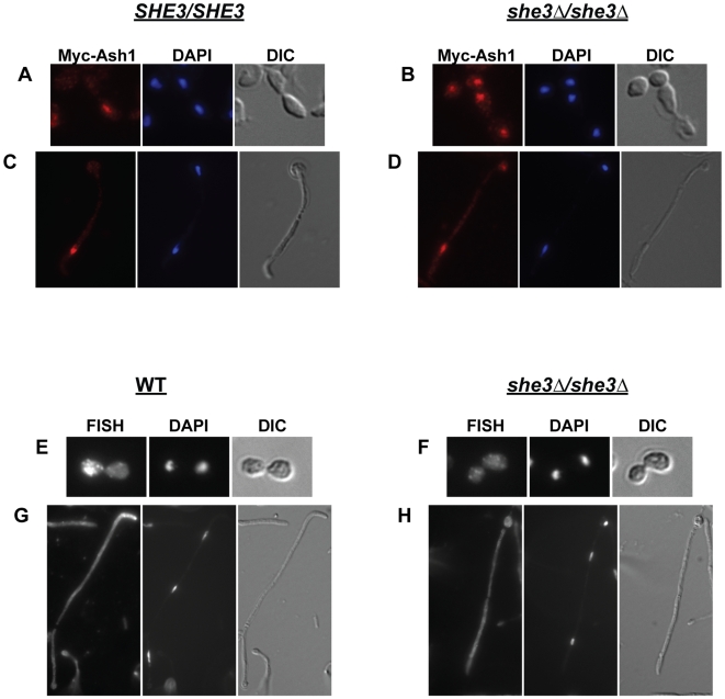
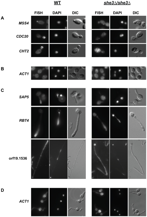
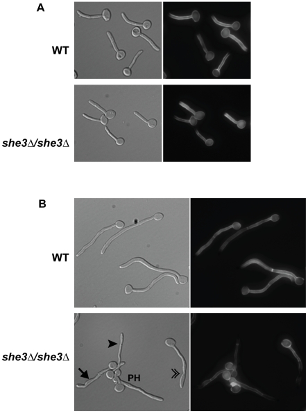
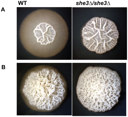
Localization of specific mRNAs is an important mechanism through which cells achieve polarity and direct asymmetric growth. Based on a framework established in Saccharomyces cerevisiae, we describe a She3-dependent RNA transport system in Candida albicans, a fungal pathogen of humans that grows as both budding (yeast) and filamentous (hyphal and pseudohyphal) forms. We identify a set of 40 mRNAs that are selectively transported to the buds of yeast-form cells and to the tips of hyphae, and we show that many of the genes encoded by these mRNAs contribute to hyphal development, as does the transport system itself. Although the basic system of mRNA transport is conserved between S. cerevisiae and C. albicans, we find that the cargo mRNAs have diverged considerably, implying that specific mRNAs can easily move in and out of transport control over evolutionary timescales. The differences in mRNA cargos likely reflect the distinct selective pressures acting on the two species.


Conflict of interest statement

The authors have declared that no competing interests exist.

Figures

Figure 1
Figure 1. Ash1 protein and ASH1 mRNA are mis-localized in C. albicans lacking She3.
(A–D) SHE3/SHE3 [SE18, (A,C)] and she3Δ/she3Δ cells [SE20, (B,D)] carrying a myc-tagged version of Ash1 (Myc-Ash1) were processed for indirect immunofluorescence, as described . Cells were stained with the mouse 9E10 anti-myc antibody followed by the Alexa-546 secondary antibody (red). Cell nuclei were visualized with DAPI (blue). In a wild type background, Myc-Ash1 accumulates in daughter cells of yeast (A) and in tip cells of hyphae (C). In the she3Δ/she3Δ strain, myc-Ash1 accumulates in both mother and daughter cells of yeast (B) and in tip and non-tip cells of hyphae (D). (E–H) Cells from wild type (“WT,” CAF2-1) and she3Δ/she3Δ (SE4) strains were processed for fluorescent in situ hybridization (FISH) to detect endogenous ASH1 transcript; cell nuclei were visualized with DAPI. Probe signal accumulates in the daughter cell of wild-type C. albicans yeast (E) and in the tips of hyphae (G). There is no specific localization of probe signal in yeast or in hyphae lacking She3 (F,H).
Figure 2
Figure 2. She3-associated transcripts accumulate in yeast buds and in hyphal tips.
Cells from wild type (“WT,” CAF2-1) and she3Δ/she3Δ (SE4) strains were processed for FISH to detect endogenous She3-associated transcripts; cell nuclei were visualized with DAPI. Representative examples illustrate She3-dependent localization of the indicated transcripts. (A) Probe signal accumulates in the incipient bud (MSS4), or bud (CDC20, CHT2) of wild type C. albicans yeast. There is no specific localization of probe signal in yeast cells lacking She3. (B) A control ACT1 probe is not localized in yeast cells from either strain. (C) In wild type hyphae, probe signal accumulates in the distal end of the germ tube (SAP5) or hyphal tip cell (RBT4, orf19.1536). Hyphae shown hybridized with SAP5, RBT4, and orf19.1536 probes were collected, respectively, 30 minutes, one hour, or three hours after serum induction. As in yeast, probe signals are not localized in hyphae lacking She3. (D) A control probe for ACT1 is not localized in germ tubes from either strain collected 40 min after serum induction. Signals from two additional control probes, ACC1 and ADH1, were not localized in yeast or hyphae (data not shown).
Figure 3
Figure 3. Morphology of C. albicans filaments from a she3 null strain.
Wild-type (“WT,” CAF2-1) and she3Δ/she3Δ (YSE4) strains were grown in YEPD/10% serum at 37°C for one (A) or two (B) hours, then fixed on cover slips and stained with Calcofluor White. (A) After one hour, germ tubes from the two strains are essentially indistinguishable. (B) By two hours, the majority of filaments from the she3-null strain display subtle defects, including swelling (→), uneven filament width (▸), and constrictions at the septal junctions in filaments that appear to have originally developed as true hyphae (»). The filament marked “PH” is a pseudohypha. The proportion of stereotypical and abnormal filaments from each strain at two hours after serum induction was determined. From 35–40 distinct fields, all fully visibly, un-branched hyphae with no constriction at junction of mother cell/filament junction were scored. Filaments displaying any of the above-described defects were scored as abnormal. Sixty-six percent of filaments from the she3-null strain (n = 167) showed some abnormality, whereas only five percent of wild type filaments (n = 171) displayed any defect.
Figure 4
Figure 4. C. albicans lacking She3 are defective in invasive growth in agar.
Wild type (“WT,” CAF2-1) and she3Δ/she3Δ (SE4) strains were grown for ten days on solid Spider medium (A) or on YEPD/10% serum medium (B). Images show representative colonies from each strain on each condition.
Figure 5
Figure 5. C. albicans She3 contributes to development of hyphae in solid media.
(A) Colonies from wild type (“WT,” CAF2-1) and she3Δ/she3Δ (SE4) strains were grown on Spider agar slabs under a glass cover slip at 30°C. Images show typical colony edges at the indicated time periods. Similar results were observed on YEPD/10% serum slabs (data not shown). (B) Colonies from wild type and she3Δ/she3Δ strains grown for five days under a cover slip placed atop a Spider agar plate.
Figure 6
Figure 6. C. albicans lacking She3 shows reduced capacity to damage oral epithelial cells.
Damage to human primary endothelial cells (A) or FaDu oral epithelial cells (B) induced by wild-type C. albicans (“WT,” QMY23), a she3Δ/SHE3 heterozygote (SE67), a she3Δ/she3Δhomozygous deletion (SE63), or a she3Δ/she3Δ strain complemented with SHE3 added to the RPS1 locus (SE64). Results for endothelial and epithelial represent the mean +/− standard deviation of, respectively, two or three independent experiments.
Figure 7
Figure 7. C. albicans strains lacking She3-associated transcripts are defective in filamentous growth.
Wild-type C. albicans (“WT,” QMY23) or strains lacking the indicated transcripts were grown for ten days on solid Spider medium at 30°C (A), on Spider agar slabs under a glass cover slip at 30°C for 48 hours (B), or in liquid YEPD with 20% serum at 37°C (C). Images are representative of two isolates of each genotype.

References

    1. Johnstone O, Lasko P. Translational regulation and RNA localization in Drosophila oocytes and embryos. Annu Rev Genet. 2001;35:365–406. - PubMed
    1. King ML, Messitt TJ, Mowry KL. Putting RNAs in the right place at the right time: RNA localization in the frog oocyte. Biol Cell. 2005;97:19–33. - PubMed
    1. Mowry KL, Cote CA. RNA sorting in Xenopus oocytes and embryos. FASEB J. 1999;13:435–445. - PubMed
    1. St Johnston D. Moving messages: the intracellular localization of mRNAs. Nat Rev Mol Cell Biol. 2005;6:363–375. - PubMed
    1. Tekotte H, Davis I. Intracellular mRNA localization: motors move messages. Trends Genet. 2002;18:636–642. - PubMed

Publication types

MeSH terms