. 2009 Sep;5(9):e1000588.
doi: 10.1371/journal.ppat.1000588. Epub 2009 Sep 25.

Bacillus anthracis lethal toxin disrupts TCR signaling in CD1d-restricted NKT cells leading to functional anergy

Affiliations

Bacillus anthracis lethal toxin disrupts TCR signaling in CD1d-restricted NKT cells leading to functional anergy

Sunil K Joshi et al. PLoS Pathog. 2009 Sep.

Erratum in

  • PLoS Pathog. 2009 Oct;5(10) doi: 10.1371/annotation/cfc9c388-05fa-4b1c-8c5f-99835278a458

Abstract

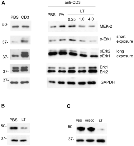
Exogenous CD1d-binding glycolipid (alpha-Galactosylceramide, alpha-GC) stimulates TCR signaling and activation of type-1 natural killer-like T (NKT) cells. Activated NKT cells play a central role in the regulation of adaptive and protective immune responses against pathogens and tumors. In the present study, we tested the effect of Bacillus anthracis lethal toxin (LT) on NKT cells both in vivo and in vitro. LT is a binary toxin known to suppress host immune responses during anthrax disease and intoxicates cells by protective antigen (PA)-mediated intracellular delivery of lethal factor (LF), a potent metalloprotease. We observed that NKT cells expressed anthrax toxin receptors (CMG-2 and TEM-8) and bound more PA than other immune cell types. A sub-lethal dose of LT administered in vivo in C57BL/6 mice decreased expression of the activation receptor NKG2D by NKT cells but not by NK cells. The in vivo administration of LT led to decreased TCR-induced cytokine secretion but did not affect TCR expression. Further analysis revealed LT-dependent inhibition of TCR-stimulated MAP kinase signaling in NKT cells attributable to LT cleavage of the MAP kinase kinase MEK-2. We propose that Bacillus anthracis-derived LT causes a novel form of functional anergy in NKT cells and therefore has potential for contributing to immune evasion by the pathogen.


Conflict of interest statement

The authors have declared that no competing interests exist.

Figures

Figure 1
Figure 1. PA binds to NKT cells.
Hepatic lymphocytes, splenocytes, lymph node cells and thymocytes were obtained from C57BL/6 mice. A Cells were stained with α-GC-loaded PE-CD1d tetramer and FITC-anti-TCRβ mAb to distinguish NKT cells, T cells and non-T/NKT cells. Samples were also counter-stained with Alexa647-PA to detect PA-binding proteins. Histograms show PA binding to each cell type. B Splenocytes were stained as described and with the concentrations of Alexa647-PA indicated. Graph shows effect of PA concentration on binding to different cell types. The histogram shows the effect of unlabeled PA on binding of Alexa647-PA. Filled histogram = no PA, continuous line = Alexa647-PA, dotted line = Alexa647-PA plus unlabeled PA. The data in A for spleen, lymph node and thymus are representative of three independent experiments. Binding of PA to hepatocytes in A and the dose response curve in B are from a single experiment. C Cell lysates were prepared from whole splenocytes (left lane) and hepatocytes (middle lane) of C57BL/6 mice (middle lane). NKT cells were enriched from the spleens of C57BL/6 mice and expanded in vitro for 5 d until 96% pure (right lane). Cell lysates were resolved by SDS-PAGE and examined by immunoblotting for the anthrax toxin receptors TEM-8 and CMG-2 and the anthrax toxin intracellular target MEK-2 which serves as a loading control.
Figure 2
Figure 2. LT alters expression of selected NKT cell surface markers.
C57BL/6 were treated with 100 µg of LT in PBS by the i.v. route or mock-treated with PBS alone. After 96 h, splenocytes were obtained and incubated with FcR-blocking mAb 2.4G2 in the presence of α-GC/CD1d tetramer, anti-TCRβ mAb and mAbs as indicated. Cells were then washed, fixed and analyzed by flow cytometry. A Shows α-GC/CD1d tetramer+/TCRβ+ NKT cells B Histograms show expression of indicated markers after gating on NKT cells. Data are representative of 3 independent experiments.
Figure 3
Figure 3. NKT cells from toxin-treated mice are viable and non-apoptotic.
C57BL/6 were treated with 100 µg of LT in PBS by the i.v. route or mock-treated with PBS alone. After 4 d, splenocytes were obtained and incubated with FcR-blocking mAb 2.4G2 in the presence of CD1d tetramer and anti-TCRβ mAb and mAb or reagents indicated. After washing and fixation, cells were analyzed by flow cytometry. Data are representative of 3 independent experiments and show uptake of 7-AAD and expression of Annexin V, PD-1 and Fas by α-GC/CD1d tetramer+/TCRβ+ cells.
Figure 4
Figure 4. LT inhibits α-GC-stimulated cytokine production.
A C57BL/6 mice were treated with 100 µg of LT in PBS by the i.v. route or mock-treated with PBS alone. After 4 d, splenocytes were obtained and stimulated in vitro with α-GC at a final concentration of 50 ng/ml. Supernatants were collected after a further 24 and 48 h and stored at −80°C. B Splenocytes from Jα18−/− mice and ex vivo-expanded NKT cells from C57BL/6 mice were treated in vitro with LT at a final concentration of 1 µg/ml for 1 h before washing and culturing separately or together in the presence or absence of α-GC as described in A. IL-4 and IFNγ concentrations in the supernatants were then determined by Bio-Plex analysis. Data show mean cytokine concentration for 3 mice per group ±SD. Asterisk indicates significant difference between cytokine concentration in un-treated control and samples from LT-treated mice. C C57BL/6 mice were treated with PBS, non-functional LT mutant, or wild type LT before enrichment of NKT cells using anti-NK1.1-based magnetic isolation. Cells were stimulated with anti-CD3 and CD28 mAbs and culture supernatants collected after 48 h.
Figure 5
Figure 5. LT inhibits TCR-mediated signaling in NKT cells.
A NKT cells were enriched from C57BL/6 splenocytes. Cells were then in vitro treated with PBS, PA (4 µg/ml) or LT (0.25 µg/ml of PA plus 0.25 µg/ml of LF), or 1.0 µg/ml or 4 µg/ml of each protein, and then stimulated by cross-linking CD3. Cell lysates were prepared and resolved by SDS-PAGE and transferred to nitrocellulose membrane before immunoblotting for MEK-2 N-terminus, phospho-Erk, total Erk, and the GAPDH loading control. Left panels show un-stimulated versus stimulated controls. Right panels show samples where CD3 was cross-linked. Two exposure times are indicated for phospho-Erk (short = 10 s, long = 90 s). B C57Bl/6 were treated with LT in vivo as described in materials and methods before collecting splenocytes and using CD1d-tetramers in conjunction with magnetic beads to obtain highly purified NKT cells. Lysates were prepared and subjected to SDS-PAGE and immunoblot analysis as indicated. C Experiment in B was repeated except that mice were treated with PBS, non-functional LT mutant, or wild type LT and NKT cells were obtained using the anti-NK1.1 enrichment method.

References

    1. Taniguchi M, Harada M, Kojo S, Nakayama T, Wakao H. The regulatory role of Valpha14 NKT cells in innate and acquired immune response. Annu Rev Immunol. 2003;21:483–513. - PubMed
    1. Galli G, Pittoni P, Tonti E, Malzone C, Uematsu Y, et al. Invariant NKT cells sustain specific B cell responses and memory. Proc Natl Acad Sci U S A. 2007;104:3984–3989. - PMC - PubMed
    1. Devera TS, Shah HB, Lang GA, Lang ML. Glycolipid-activated NKT cells support the induction of persistent plasma cell responses and antibody titers. Eur J Immunol. 2008;38:1001–1011. - PMC - PubMed
    1. Hermans IF, Silk JD, Gileadi U, Salio M, Mathew B, et al. NKT cells enhance CD4+ and CD8+ T cell responses to soluble antigen in vivo through direct interaction with dendritic cells. J Immunol. 2003;171:5140–5147. - PubMed
    1. Lang GA, Devera TS, Lang ML. Requirement for CD1d expression by B cells to stimulate NKT cell-enhanced antibody production. Blood. 2008;111:2158–2162. - PMC - PubMed

Publication types

MeSH terms