. 2009 Sep;5(9):e1000591.
doi: 10.1371/journal.ppat.1000591. Epub 2009 Sep 25.

Ion-abrasion scanning electron microscopy reveals surface-connected tubular conduits in HIV-infected macrophages

Affiliations

Ion-abrasion scanning electron microscopy reveals surface-connected tubular conduits in HIV-infected macrophages

Adam E Bennett et al. PLoS Pathog. 2009 Sep.

Abstract

HIV-1-containing internal compartments are readily detected in images of thin sections from infected cells using conventional transmission electron microscopy, but the origin, connectivity, and 3D distribution of these compartments has remained controversial. Here, we report the 3D distribution of viruses in HIV-1-infected primary human macrophages using cryo-electron tomography and ion-abrasion scanning electron microscopy (IA-SEM), a recently developed approach for nanoscale 3D imaging of whole cells. Using IA-SEM, we show the presence of an extensive network of HIV-1-containing tubular compartments in infected macrophages, with diameters of approximately 150-200 nm, and lengths of up to approximately 5 microm that extend to the cell surface from vesicular compartments that contain assembling HIV-1 virions. These types of surface-connected tubular compartments are not observed in T cells infected with the 29/31 KE Gag-matrix mutant where the virus is targeted to multi-vesicular bodies and released into the extracellular medium. IA-SEM imaging also allows visualization of large sheet-like structures that extend outward from the surfaces of macrophages, which may bend and fold back to allow continual creation of viral compartments and virion-lined channels. This potential mechanism for efficient virus trafficking between the cell surface and interior may represent a subversion of pre-existing vesicular machinery for antigen capture, processing, sequestration, and presentation.


Conflict of interest statement

The authors have declared that no competing interests exist.

Figures

Figure 1
Figure 1. Cryo-electron tomography of HIV-1 infected monocyte-derived macrophages (MDM).
(A) Projection image of the edge of an infected cell at low magnifications indicating that no useful image contrast is obtained in most regions of the cell except at the very outer edges. (B) Projection image at higher magnification near the edge of the cell showing filopodial extensions and clusters of virions in close proximity. The inset shows a magnified view of individual filopodia, denoised to enhance visualization of the actin bundles that make up the interior. The small black dots that are especially noticeable in panel (B) represent 15 nm-sized gold particles added to the grid for purposes of providing fiducials for tomographic reconstruction. Scale bars: panel (A) 1 µm, panel (B) and inset 200 nm.
Figure 2
Figure 2. Electron tomography of a 150 nm thick section from HIV-1 BaL infected macrophages.
A tomographic slice (nominal thickness 1 nm) through a region of the cell containing a collection of viruses in an internal compartment. The expanded insets show zoomed-in images of individual immature (top) and mature (bottom) virions. Scale bar 100 nm.
Figure 3
Figure 3. Virion reservoirs and channels in primary HIV-1-infected MDM revealed by IA-SEM imaging.
(A) Single cross-sectional image shows internal compartments, highlighted further in (B) indicating budding (light blue arrow), immature (red arrow), and mature (yellow arrow) virions, and membrane boundary (pink arrow). (C) Some compartments deep in the interior are seen to display “bagpipe”-like protrusions that are filled with virions. Scale bars are 200 nm long in panels (A)–(C). (D) Segmented 3D image illustrating the distribution of virions (red) within a reservoir connected to the plasma membrane. A 2D scale bar cannot be used for the 3D perspective rendering in (D); for reference, however, each virion has an approximate diameter of 120 nm.
Figure 4
Figure 4. Virion channels of roughly uniform diameter allow communication between deep internal reservoirs and the plasma membrane of primary HIV-1-infected MDM.
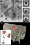
Individual IA-SEM images of virion channels identified by dual beam imaging are shown in (A) transverse and (B–D) axial sections. Scale bars are 100 nm long. (E) and inset: Illustration of the depth of some of the virion channels by automated segmentation of the raw 3D image (shown in Video S2) of a portion of an HIV-1 infected macrophage with two internal compartments, each of which contains numerous virions (red) with an approximate diameter of ∼120 nm. Virion channels connecting to the surface are observed from the compartment on the left, while the compartment on the right is completely internal. An animation of the segmented volume is included in Video S7.
Figure 5
Figure 5. Transmission electron microscopic and IA-SEM imaging of Jurkat T cells infected with the VSV-G pseudotyped 29/31 HIV-1 Gag matrix mutant.
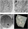
(A, B) Selected projection TEM images from a 100 nm thick section obtained from fixed, osmium-stained, plastic-embedded cells. Small vacuolar compartments, some containing viruses can be visualized in the interior of the cell. (C) A single IA-SEM image from the interior of the same block used to obtain the TEM images in panel (A). A complete stack of images showing the distribution and closed shapes of the compartments through this and another cell is included in Videos S8 and S9. (D) Rendering of the membrane conpartments in the interior of the cell illustrating that they are closed, and not connected to the cell surface irrespective of whether or not they appear to contain viruses (red). Scale bars in all panels are 1 µm wide.
Figure 6
Figure 6. 3D representation of the surface and interior of an HIV-infected macrophage (animation is presented in Video S10).
(A) Sections that would appear to contain “filopodia” when imaged by transmission electron microscopy of individual sections can actually correspond to large wavelike membrane processes as in this example. The virions are shown in red. (B, C) Schematic side (B) and front (C) views of these surface protrusions shown to indicate how bending and folding back of the extensions onto the surface of the cell could trap the contents of the aqueous environment within the invaginated folds of the membrane, and allow creation of viral compartments.

References

    1. Orenstein JM. Isn't a picture still worth a thousand words? Ultrastruct Pathol. 2000;24:67–74. - PubMed
    1. Heymann JA, Hayles M, Gestmann I, Giannuzzi LA, Lich B, et al. Site-specific 3D imaging of cells and tissues with a dual beam microscope. J Struct Biol. 2006;155:63–73. - PMC - PubMed
    1. Heymann JA, Shi D, Kim S, Bliss D, Milne JL, et al. 3D imaging of mammalian cells with ion-abrasion scanning electron microscopy. J Struct Biol. 2009;166:1–7. - PMC - PubMed
    1. Hildebrand M, Kim S, Shi D, Scott K, Subramaniam S. 3D imaging of diatoms with ion-abrasion scanning electron microscopy. J Struct Biol. 2009;166:316–328. - PMC - PubMed
    1. Gousset K, Ablan SD, Coren LV, Ono A, Soheilian F, et al. Real-time visualization of HIV-1 GAG trafficking in infected macrophages. PLoS Pathog. 2008;4:e1000015. doi: 10.1371/journal.ppat.1000015. - DOI - PMC - PubMed

Publication types