. 2009 Sep 25;4(9):e7204.
doi: 10.1371/journal.pone.0007204.

Panton-valentine leukocidin enhances the severity of community-associated methicillin-resistant Staphylococcus aureus rabbit osteomyelitis

Affiliations

Panton-valentine leukocidin enhances the severity of community-associated methicillin-resistant Staphylococcus aureus rabbit osteomyelitis

Anne-Claude Crémieux et al. PLoS One. .

Abstract

Background: Extensive spread of community-associated methicillin-resistant Staphylococcus aureus (CA-MRSA) in the United States, and the concomitant increase in severe invasive staphylococcal infections, including osteomyelitis, in healthy children, has led to renewed interest in Panton-Valentine leukocidin (PVL). However, the pathogenetic role of PVL in staphylococcal infections remains controversial, possibly because it depends on the site of infection.

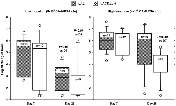
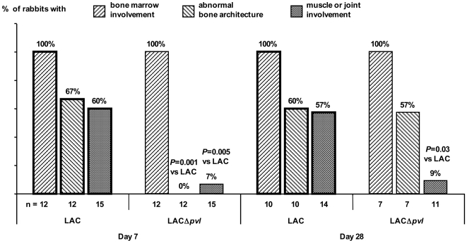
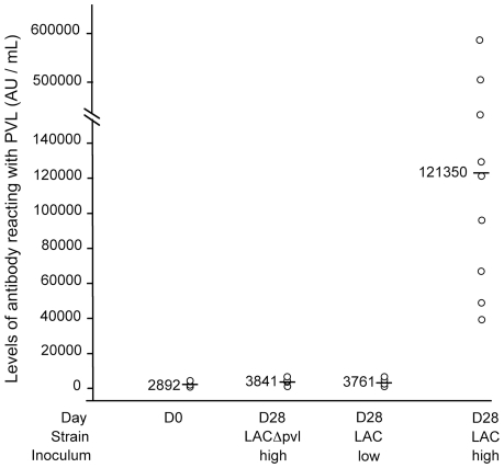
Methodology/principal findings: We compared the course of experimental rabbit osteomyelitis due to the PVL-positive CA-MRSA strain USA 300 (LAC) and its PVL-negative isogenic derivative (LACDeltapvl), using a low and a high inoculum (8x10(5) and 4x10(8) CFU). With the low inoculum, bone infection was less frequent on day 7 (D7) and day 28 (D28) with LACDeltapvl than with LAC (respectively 12/19 and 18/19 animals, p = 0.042). With the high inoculum of both strains, all the animals were infected on D7 and the infection persisted on D28 in almost every case. However, tibial bacterial counts and the serum CRP concentration fell significantly between D7 and D28 with LACDeltapvl but not with LAC. Respectively 67% and 60% of LAC-infected rabbits had bone deformation and muscle/joint involvement on D7, compared to 0% and 7% of LACDeltapvl-infected rabbits (p = 0.001 and p = 0.005 respectively). Between D0 and D28, the anti-PVL antibody titer increased significantly only with the high inoculum of LAC.

Conclusions/significance: PVL appears to play a role in the persistence and rapid local extension of rabbit osteomyelitis, in keeping with the greater severity of human bone infections due to PVL-positive S. aureus. The possible therapeutic implications of these findings are discussed.


Conflict of interest statement

Competing Interests: The authors have declared that no competing interests exist.

Figures

Figure 1
Figure 1. Bacterial counts in rabbit tibia.
Box plot representation (10, 25, 50, 75, 90th percentiles and outliers) of bacterial counts in rabbit tibia 7 days (D7) and 28 days (D28) after inoculation with 8×10⁵ (low) or 4×10⁸ (high) CFU of LAC and LAC Δpvl CA-MRSA. P values for differences between D28 and D7 were obtained with the non parametric Mann-Whitney U-test.
Figure 2
Figure 2. Serial MRI of a rabbit challenged with a low inoculum of LAC (PVL+) or LACΔpvl (PVL−).
2A - Serial magnetic resonance imaging (MRI) of a rabbit infected with a low inoculum of LAC (PVL+); the mean tibial bacterial count was 4.64 Log10 CFU/g of bone on D28. The infection appears as a marrow hypersignal on D7, that becomes more intense and extends to the entire diaphysis on days 14 and 21. A medullary abscess was found at autopsy on D28. 2B - Serial MRI of a rabbit challenged with a low inoculum of LACΔpvl (PVL−). An intramedullary abscess was visible in the metaphysis and proximal diaphysis on D7, appearing as an intense marrow hypersignal. Central necrosis was noted on images obtained on D14 and D21. Bone marrow necrosis was observed macroscopically on D28. The mean tibial bacterial count on D28 was 6.08 Log10 CFU/g of bone.
Figure 3
Figure 3. Bone and muscle/joint involvement after challenge with a high inoculum of LAC and LAC Δpvl.
Percentages of rabbits with bone marrow involvement, bone deformation and muscle/joint involvement 7 (D7) and 28 (D28) days after challenge with a high inoculum of LAC and LAC Δpvl. n is the total number of animals. P values for differences between the c and LAC groups were obtained with the non parametric Mann-Whitney U-test.
Figure 4
Figure 4. Typical radiological and histological findings after challenge with a high inoculum of LAC (PVL+).
A, B, C: Radiological and histological findings 28 days after challenge with a high inoculum of LAC (PVL+). Note the major deformation and widening of the entire diaphysis, compared to the control animal injected with sclerosing agent alone. Histological studies showed an intramedullary abscess and signs of chronic osteomyelitis. B: Bone abscess: the bone marrow space is filled with altered neutrophilic PMN, accompanying bone destruction and necrosis. C: Sequestrum: area of necrotic bone surrounded by an acute inflammatory exudate (pus). D, E, F: 28 days after challenge with a high inoculum of LAC (PVL+), note the soft tissue abscess surrounded by a fibrous layer on MRI and histological studies. G, H, I: Histological findings in a rabbit (PVL+) that died on D8 after challenged with a high inoculum of LAC. Histological sections (H) show muscle involvement with diffuse necrosis and dystrophic calcification, and (I) a purulent exudate in the joint cavity.
Figure 5
Figure 5. Anti-PVL antibody levels in sera of rabbits.
Distribution of anti-PVL antibody levels in 24 sera obtained before S. aureus challenge (D0); 7 sera obtained 28 days after challenge with LACΔpvl, 9 sera obtained 28 days after challenge with a high inoculum of LAC, and 8 sera obtained 28 days after challenge with a low inoculum of LAC. Bars show the median values. The median anti-PVL titer increased 31-fold on D28 versus D0 after challenge with a high inoculum of LAC (p<0.001). No significant change was detected at D28 versus D0 in animals challenged with LACΔpvl or with the low inoculum of LAC.
Figure 6
Figure 6. C-reactive protein (CRP) values in rabbits.
Distribution of C-reactive protein (CRP) values in 36 rabbits (9 samples obtained on D7 from rabbits infected with LAC, 9 samples obtained on D28 from rabbits infected with LACΔpvl, 9 samples obtained on D28 from rabbits infected with LAC, and 9 samples obtained on D28 from rabbits infected with LACΔpvl (all animals challenged with the high inoculum). Bars show the median values. The fall in the median CRP values from D7 to D28 was statistically significant for LACΔpvl infection (p = 002), but not for LAC infection (p = .085).

References

    1. Tristan A, Bes M, Meugnier H, Lina G, Bozdogan B, et al. Global distribution of Panton-Valentine leukocidin–positive methicillin-resistant Staphylococcus aureus, 2006. Emerg Infect Dis. 2007;13:594–600. - PMC - PubMed
    1. Lina G, Piemont Y, Godail-Gamot F, Bes M, Peter MO, et al. Involvement of Panton-Valentine leukocidin-producing Staphylococcus aureus in primary skin infections and pneumonia. Clin Infect Dis. 1999;29:1128–1132. - PubMed
    1. Gillet Y, Issartel B, Vanhems P, Fournet JC, Lina G, et al. Association between Staphylococcus aureus strains carrying gene for Panton-Valentine leukocidin and highly lethal necrotising pneumonia in young immunocompetent patients. Lancet. 2002;359:753–759. - PubMed
    1. Gillet Y, Vanhems P, Lina G, Bes M, Vandenesch F, et al. Factors associated with the high fatality rate of Panton Valentine Leukocidin associated Staphylococcus aureus necrotising pneumonia. Clin Infect Dis. 2007;45:315–321. - PubMed
    1. Dohin B, Gillet Y, Kohler R, Lina G, Vandenesch F, et al. Pediatric bone and joint infections caused by Panton-Valentine leukocidin-positive Staphylococcus aureus. Pediatr Infect Dis J. 2007;26:1042–1048. - PubMed

Publication types

MeSH terms