

Modeling familial British and Danish dementia
- PMID: 19779737
- PMCID: PMC8375673
- DOI: 10.1007/s00429-009-0221-9
Modeling familial British and Danish dementia
Abstract
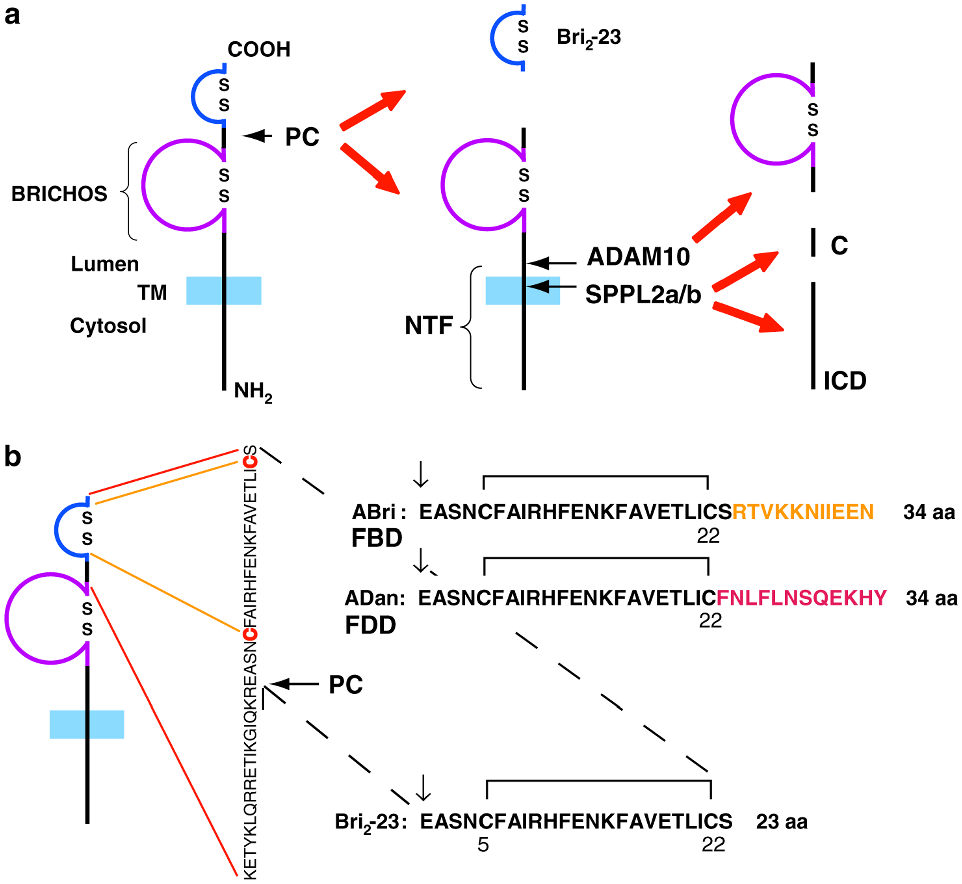
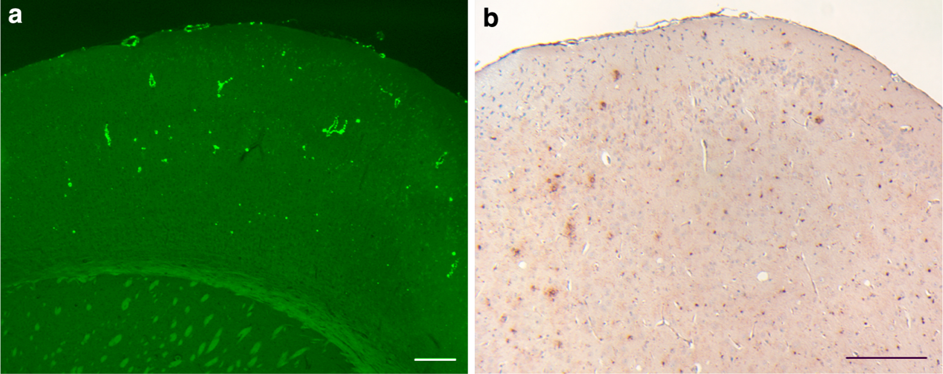
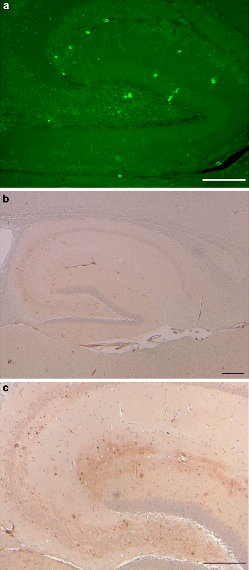
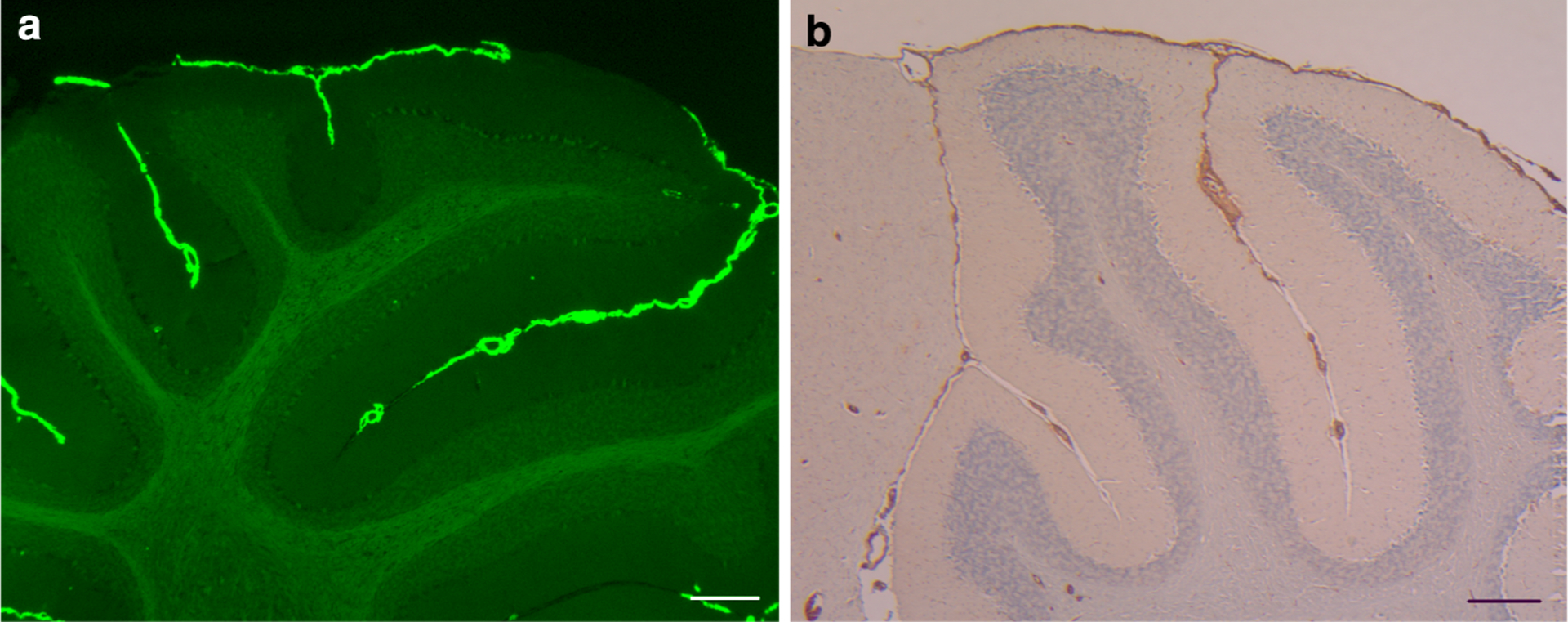
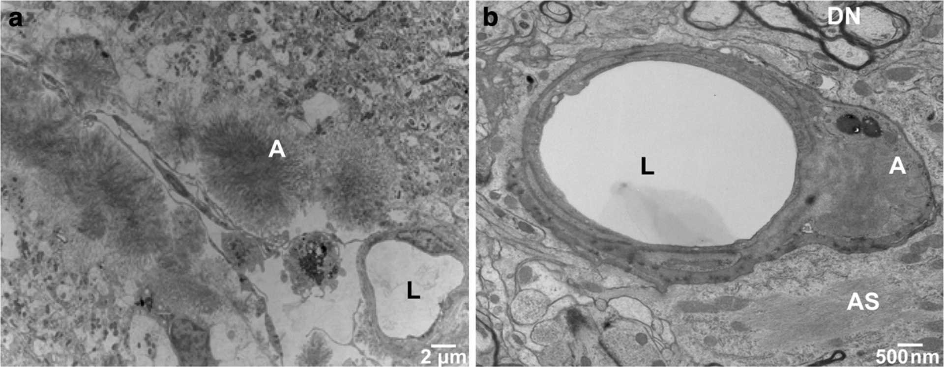
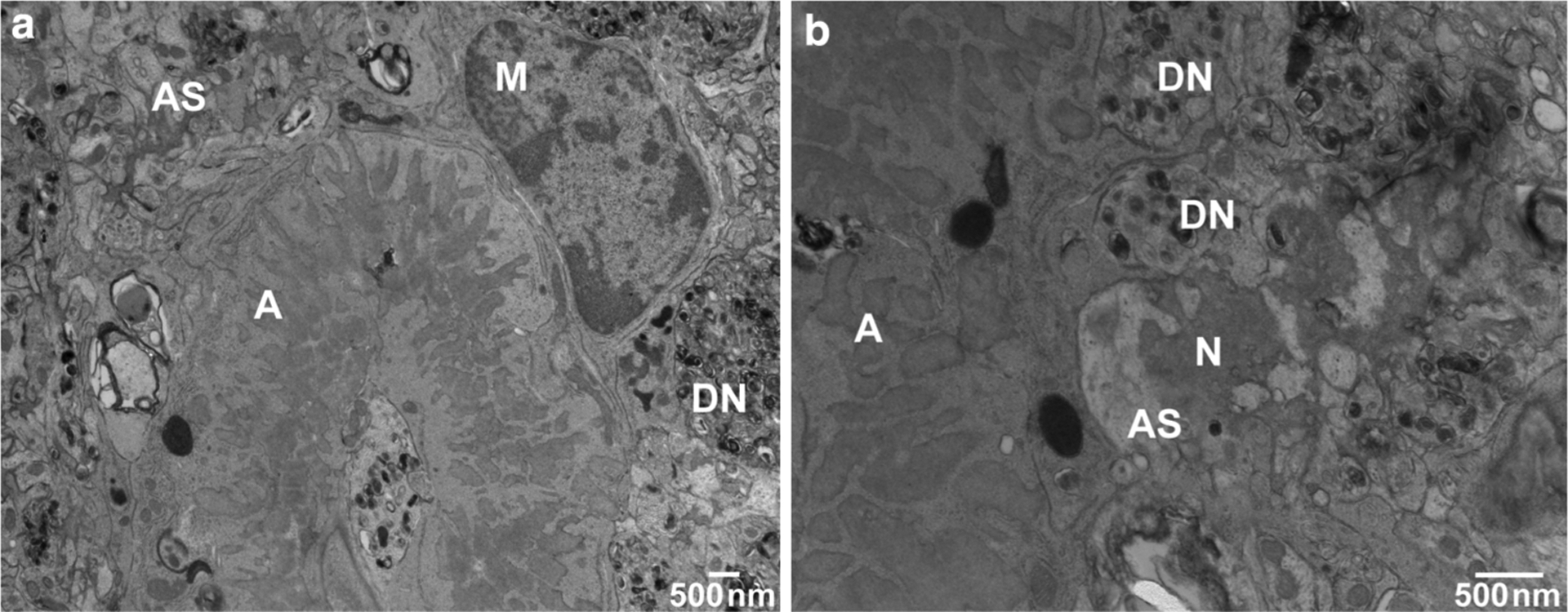
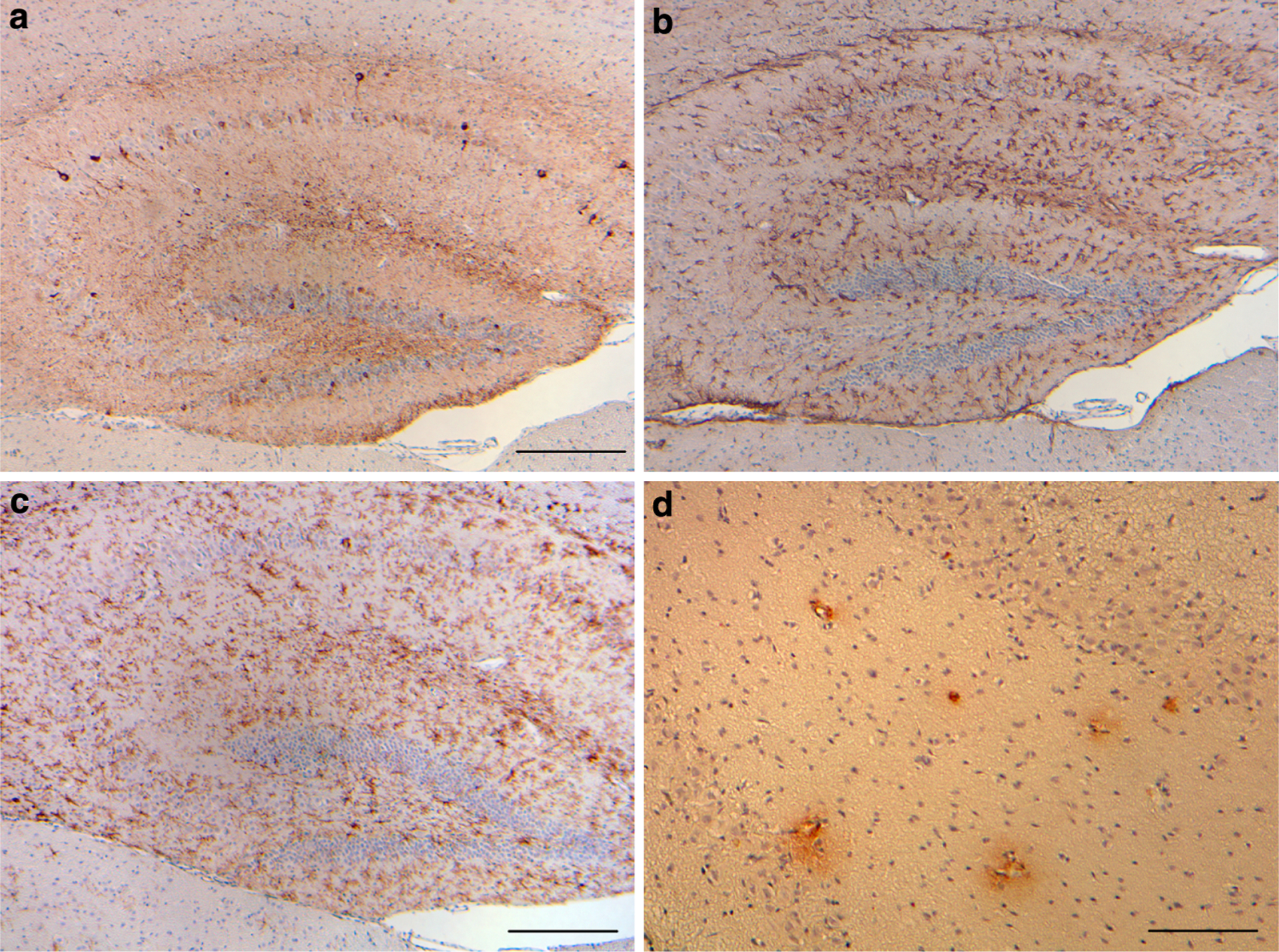
Familial British dementia (FBD) and familial Danish dementia (FDD) are two autosomal dominant neurodegenerative diseases caused by mutations in the BRI ( 2 ) gene. FBD and FDD are characterized by widespread cerebral amyloid angiopathy (CAA), parenchymal amyloid deposition, and neurofibrillary tangles. Transgenic mice expressing wild-type and mutant forms of the BRI(2) protein, Bri ( 2 ) knock-in mutant mice, and Bri ( 2 ) gene knock-out mice have been developed. Transgenic mice expressing a human FDD-mutated form of the BRI ( 2 ) gene have partially reproduced the neuropathological lesions observed in FDD. These mice develop extensive CAA, parenchymal amyloid deposition, and neuroinflammation in the central nervous system. These animal models allow the study of the molecular mechanism(s) underlying the neuronal dysfunction in these diseases and allow the development of potential therapeutic approaches for these and related neurodegenerative conditions. In this review, a comprehensive account of the advances in the development of animal models for FBD and FDD and of their relevance to the study of Alzheimer disease is presented.
Figures
Similar articles
-
Cerebral amyloid angiopathy and parenchymal amyloid deposition in transgenic mice expressing the Danish mutant form of human BRI2.Brain Pathol. 2009 Jan;19(1):58-68. doi: 10.1111/j.1750-3639.2008.00164.x. Epub 2008 Apr 10. Brain Pathol. 2009. PMID: 18410407 Free PMC article.
-
Chemical traits of cerebral amyloid angiopathy in familial British-, Danish-, and non-Alzheimer's dementias.J Neurochem. 2022 Nov;163(3):233-246. doi: 10.1111/jnc.15694. Epub 2022 Sep 25. J Neurochem. 2022. PMID: 36102248 Free PMC article.
-
Memory deficits due to familial British dementia BRI2 mutation are caused by loss of BRI2 function rather than amyloidosis.J Neurosci. 2010 Nov 3;30(44):14915-24. doi: 10.1523/JNEUROSCI.3917-10.2010. J Neurosci. 2010. PMID: 21048150 Free PMC article.
-
Modeling familial British dementia in transgenic mice.Brain Pathol. 2006 Jan;16(1):80-5. doi: 10.1111/j.1750-3639.2006.tb00564.x. Brain Pathol. 2006. PMID: 16612985 Free PMC article. Review.
-
Preamyloid lesions and cerebrovascular deposits in the mechanism of dementia: lessons from non-beta-amyloid cerebral amyloidosis.Neurodegener Dis. 2008;5(3-4):173-5. doi: 10.1159/000113694. Epub 2008 Mar 6. Neurodegener Dis. 2008. PMID: 18322382 Free PMC article. Review.
Cited by
-
Increased tau phosphorylation and tau truncation, and decreased synaptophysin levels in mutant BRI2/tau transgenic mice.PLoS One. 2013;8(2):e56426. doi: 10.1371/journal.pone.0056426. Epub 2013 Feb 13. PLoS One. 2013. PMID: 23418567 Free PMC article.
-
Functional BRI2-TREM2 interactions in microglia: implications for Alzheimer's and related dementias.EMBO Rep. 2024 Mar;25(3):1326-1360. doi: 10.1038/s44319-024-00077-x. Epub 2024 Feb 12. EMBO Rep. 2024. PMID: 38347225 Free PMC article.
-
Updated Outlook of Cerebral Amyloid Angiopathy and Inflammatory Subtypes: Pathophysiology, Clinical Manifestations, Diagnosis and Management.J Alzheimers Dis Rep. 2022 Oct 18;6(1):627-639. doi: 10.3233/ADR-220055. eCollection 2022. J Alzheimers Dis Rep. 2022. PMID: 36447738 Free PMC article. Review.
-
The role of the integral type II transmembrane protein BRI2 in health and disease.Cell Mol Life Sci. 2021 Nov;78(21-22):6807-6822. doi: 10.1007/s00018-021-03932-5. Epub 2021 Sep 4. Cell Mol Life Sci. 2021. PMID: 34480585 Free PMC article. Review.
-
APP heterozygosity averts memory deficit in knockin mice expressing the Danish dementia BRI2 mutant.EMBO J. 2011 May 17;30(12):2501-9. doi: 10.1038/emboj.2011.161. EMBO J. 2011. PMID: 21587206 Free PMC article.
References
-
- Aigner L, Arber S, Kapfhammer JP, Laux T, Schneider C, Botteri F, Brenner HR, Caroni P (1995) Overexpression of the neural growth-associated protein GAP-43 induces nerve sprouting in the adult nervous system of transgenic mice. Cell 83:269–278 - PubMed
-
- Akiyama H, Kondo H, Arai T, Ikeda K, Kato M, Iseki E, Schwab C, McGeer PL (2004) Expression of BRI, the normal precursor of the amyloid protein of familial British dementia, in human brain. Acta Neuropathol 107:53–58 - PubMed
-
- Borchelt DR, Davis J, Fischer M, Lee MK, Slunt HH, Ratovitsky T, Regard J, Copeland NG, Jenkins NA, Sisodia SS, Price DL (1996) A vector for expressing foreign genes in the brains and hearts of transgenic mice. Genet Anal 13:159–163 - PubMed
-
- Choi SI, Vidal R, Frangione B, Levy E (2004) Axonal transport of British and Danish amyloid peptides via secretory vesicles. FASEB J 18:373–375 - PubMed
-
- Coomaraswamy J, Herzig MC, Kaeser SA, Ghiso J, Jucker M (2006) Transgenic mouse models of familial British and Danish dementias. ICAD S116:P1–P072
Publication types
MeSH terms
Substances
Grants and funding
LinkOut - more resources
Full Text Sources
Other Literature Sources
Medical
