

Changes in global gene expression during in vitro decidualization of rat endometrial stromal cells
- PMID: 19780023
- PMCID: PMC2840620
- DOI: 10.1002/jcp.21929
Changes in global gene expression during in vitro decidualization of rat endometrial stromal cells
Abstract
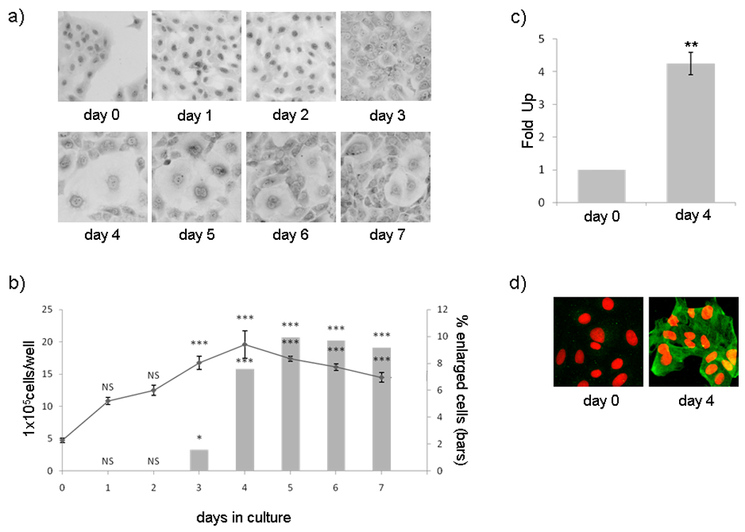
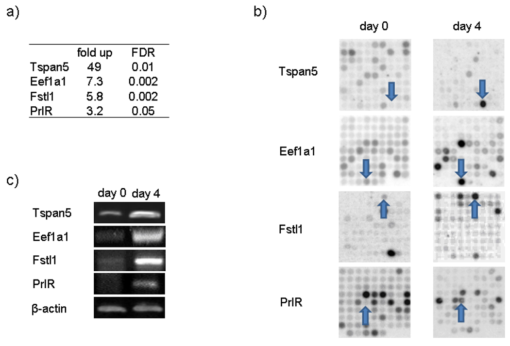
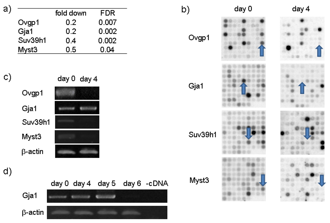
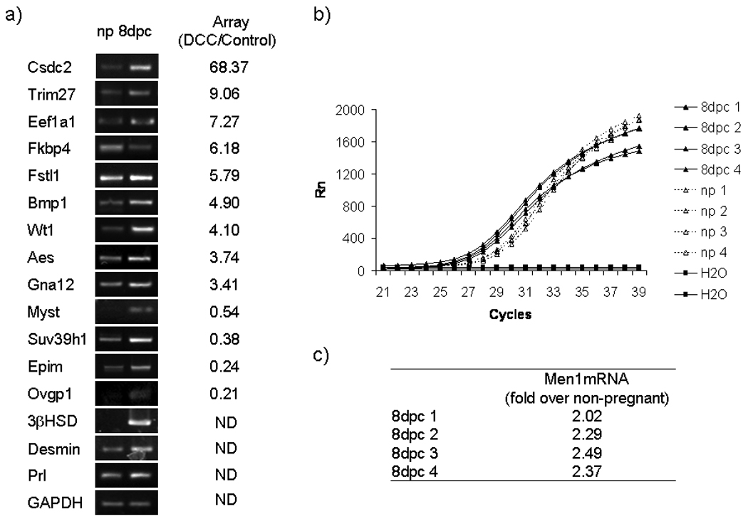
During the preimplantation phase of pregnancy the endometrial stroma differentiates into decidua, a process that implies numerous morphological changes and is an example of physiological transdifferentiation. Here we show that UIII rat endometrial stromal cells cultured in the presence of calf serum acquired morphological features of decidual cells and expressed decidual markers. To identify genes involved in decidualization we compared gene expression patterns of control and decidualized UIII cells using cDNA microarray. We found 322 annotated genes exhibiting significant differences in expression (>3-fold, fold discovery rate (FDR) >0.005), of which 312 have not been previously related to decidualization. Analysis of overrepresented functions revealed that protein synthesis, gene expression, and chromatin architecture and remodeling are the most relevant modified functions during decidualization. Relevant genes are also found in the functional terms differentiation, cell proliferation, signal transduction, and matrix/structural proteins. Several of these new genes involved in decidualization (Csdc2, Trim27, Eef1a1, Bmp1, Wt1, Aes, Gna12, and Men1) are shown to be also regulated in uterine decidua during normal pregnancy. Thus, the UIII cell culture model will allow future mechanistic studies to define the transcriptional network regulating reprogramming of stromal cells into decidual cells.
Figures
Similar articles
-
miRNA signature and Dicer requirement during human endometrial stromal decidualization in vitro.PLoS One. 2012;7(7):e41080. doi: 10.1371/journal.pone.0041080. Epub 2012 Jul 20. PLoS One. 2012. PMID: 22911744 Free PMC article.
-
MicroRNA-145 targets Smad1 in endometrial stromal cells and regulates decidualization in rat.J Mol Med (Berl). 2019 Apr;97(4):509-522. doi: 10.1007/s00109-019-01744-6. Epub 2019 Feb 7. J Mol Med (Berl). 2019. PMID: 30729278
-
Growth regulation by estrogen in breast cancer 1 (GREB1) is a novel progesterone-responsive gene required for human endometrial stromal decidualization.Mol Hum Reprod. 2017 Sep 1;23(9):646-653. doi: 10.1093/molehr/gax045. Mol Hum Reprod. 2017. PMID: 28911214 Free PMC article.
-
Shp2 in uterine stromal cells critically regulates on time embryo implantation and stromal decidualization by multiple pathways during early pregnancy.PLoS Genet. 2022 Jan 13;18(1):e1010018. doi: 10.1371/journal.pgen.1010018. eCollection 2022 Jan. PLoS Genet. 2022. PMID: 35025868 Free PMC article.
-
Epigenetic modifications working in the decidualization and endometrial receptivity.Cell Mol Life Sci. 2020 Jun;77(11):2091-2101. doi: 10.1007/s00018-019-03395-9. Epub 2019 Dec 7. Cell Mol Life Sci. 2020. PMID: 31813015 Free PMC article. Review.
Cited by
-
Exploring the role of Luman/CREB3 in regulating decidualization of mice endometrial stromal cells by comparative transcriptomics.BMC Genomics. 2020 Jan 30;21(1):103. doi: 10.1186/s12864-020-6515-2. BMC Genomics. 2020. PMID: 32000663 Free PMC article.
-
Role of Csdc2 in Regulating Secondary Hair Follicle Growth in Cashmere Goats.Int J Mol Sci. 2024 Jul 30;25(15):8349. doi: 10.3390/ijms25158349. Int J Mol Sci. 2024. PMID: 39125915 Free PMC article.
-
Tumor Necrosis Factor Alpha and Insulin-Like Growth Factor 1 Induced Modifications of the Gene Expression Kinetics of Differentiating Skeletal Muscle Cells.PLoS One. 2015 Oct 8;10(10):e0139520. doi: 10.1371/journal.pone.0139520. eCollection 2015. PLoS One. 2015. PMID: 26447881 Free PMC article.
-
CDC2 mediates progestin initiated endometrial stromal cell proliferation: a PR signaling to gene expression independently of its binding to chromatin.PLoS One. 2014 May 23;9(5):e97311. doi: 10.1371/journal.pone.0097311. eCollection 2014. PLoS One. 2014. PMID: 24859236 Free PMC article.
-
Crude bark extract of Dysozylum alliarium induces alteration in histological structures and VEGF-C expression in uterus during days 4-7 of gestation in albino rat.Reprod Med Biol. 2013 Feb 28;12(3):85-98. doi: 10.1007/s12522-012-0143-8. eCollection 2013 Jul. Reprod Med Biol. 2013. PMID: 29699135 Free PMC article.
References
-
- Agarwal SK, Guru SC, Heppner C, Erdos MR, Collins RM, Park SY, Saggar S, Chandrasekharappa SC, Collins FS, Spiegel AM, Marx SJ, Burns AL. Menin Interacts with the AP1 Transcription Factor JunD and Represses JunD-Activated Transcription. Cell. 1999;96:143–152. - PubMed
-
- Arai KY, Tsuchida K, Uehara K, Taya K, Sugino H. Characterization of rat follistatin-related gene: effects of estrous cycle stage and pregnancy on its messenger RNA expression in rat reproductive tissues. Biol Reprod. 2003;68(1):199–206. - PubMed
-
- Bany BM, Zhang X, Kennedy TG. Effects of epidermal growth factor and interleukin-1alpha on plasminogen activator secretion and decidualization in rat endometrial stromal cells. Biol Reprod. 1998;59(1):131–135. - PubMed
-
- Beissbarth T, Speed TP. GOstat: find statistically overrepresented Gene Ontologies within a group of genes. Bioinformatics. 2004;20(9):1464–1465. - PubMed
-
- Benjamini Y, Hochberg Y. Controlling the False Discovery Rate: A Practical and Powerful Approach to Multiple Testing. J Roy Stat Soc B. 1995;57:289–300.
Publication types
MeSH terms
Grants and funding
LinkOut - more resources
Full Text Sources
Molecular Biology Databases
Miscellaneous
