. 2009 Nov;62(5):1221-31.
doi: 10.1002/mrm.22115.

Independent slab-phase modulation combined with parallel imaging in bilateral breast MRI

Affiliations

Independent slab-phase modulation combined with parallel imaging in bilateral breast MRI

Misung Han et al. Magn Reson Med. 2009 Nov.

Abstract

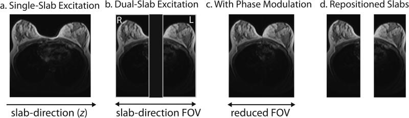
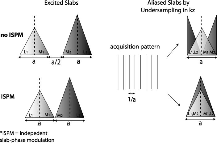
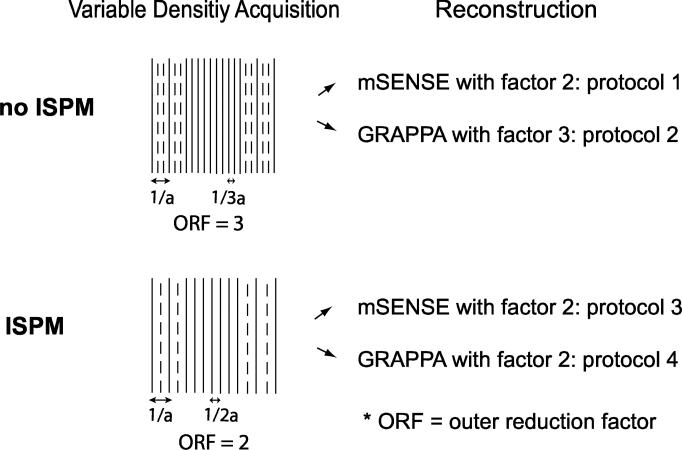
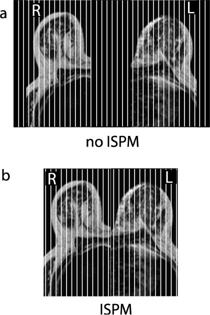
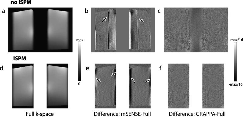
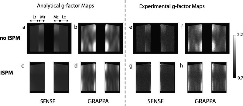
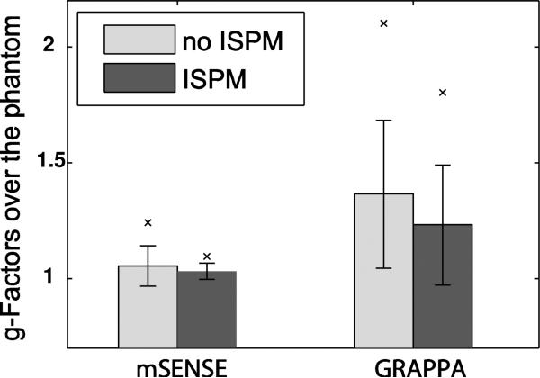
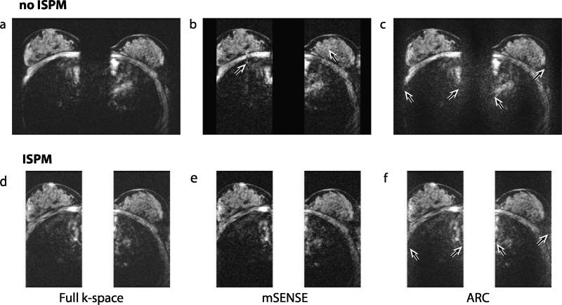
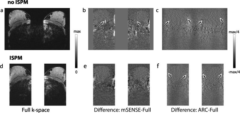
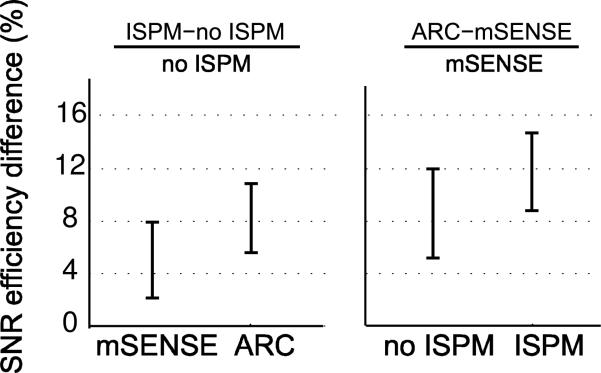
Independent slab-phase modulation allows three-dimensional imaging of multiple volumes without encoding the space between volumes, thus reducing scan time. Parallel imaging further accelerates data acquisition by exploiting coil sensitivity differences between volumes. This work compared bilateral breast image quality from self-calibrated parallel imaging reconstruction methods such as modified sensitivity encoding, generalized autocalibrating partially parallel acquisitions and autocalibrated reconstruction for Cartesian sampling (ARC) for data with and without slab-phase modulation. A study showed an improvement of image quality by incorporating slab-phase modulation. Geometry factors measured from phantom images were more homogenous and lower on average when slab-phase modulation was used for both mSENSE and GRAPPA reconstructions. The resulting improved signal-to-noise ratio (SNR) was validated for in vivo images as well using ARC instead of GRAPPA, illustrating average SNR efficiency increases in mSENSE by 5% and ARC by 8% based on region of interest analysis. Furthermore, aliasing artifacts from mSENSE reconstruction were reduced when slab-phase modulation was used. Overall, slab-phase modulation with parallel imaging improved image quality and efficiency for 3D bilateral breast imaging.


Figures

Figure 1
Figure 1
Axial views showing sagittal slab positions for standard excitation, dual-slab excitation and independent slab-phase modulation. (a) Single-slab excitation excites a large volume including the right and left breasts and the space between them. (b) A dual-band pulse excites a slab over each breast simultaneously but independently; however, for simultaneous imaging of the two slabs, the two excited slabs and the space between them must be phase-encoded together. (c) By incorporating slab-phase modulation to the dual-band pulse, the two slabs are virtually shifted close together and the slab-direction FOV can be reduced. (d) After image reconstruction, the two slabs can be repositioned if the physical slab locations are known.
Figure 2
Figure 2
Slab-phase modulation and undersampling. If two slabs with each thickness of a are separated by the distance of a/2, the total slab-direction FOV is 2.5a but by incorporating ISPM, the required FOV can be reduced to 2a. If undersampling is conducted in kz with the sampling interval of 1/a, two slabs are superimposed. When ISPM is not used, the center of the two slabs are misaligned, thus the medial region of one slab (M1) overlaps with the medial region of the other slab (M2), and the lateral region of one slab (L1) overlaps with the lateral region of other slab (L2). When ISPM is incorporated, the centers of the two slabs are aligned and the lateral region of one slab overlaps with the medial region of the other slab. The use of ISPM maintains the constant actual separation between superimposed slices, whereas if ISPM is not used, the separation varies.
Figure 3
Figure 3
Self-calibrated parallel imaging and reconstruction protocols. To apply self-calibrated parallel imaging techniques, extra central kz planes are acquired in addition to undersampled acquisition with interval 1/a. When ISPM is not used, central kz planes are acquired with interval 1/3a for adequate coil sensitivity information; however when ISPM is used, the central kz planes are acquired with interval 1/2a. So the outer reduction factors are 3 and 2 for each case. For reconstruction, the data without ISPM is reconstructed using mSENSE with a factor of 2 and GRAPPA with a factor of 3 (protocol 1,2). The data with ISPM is reconstructed using mSENSE with a factor of 2 and GRAPPA with a factor of 2 (protocol 3,4).
Figure 4
Figure 4
Slab positions, phase-encoded sections and FOV. (a) When ISPM was not applied, the two excited slabs and the empty space between them were phase-encoded with 192 sagittal 3D sections. (b) When ISPM was applied, the two excited slabs were encoded with 128 sagittal 3D sections.
Figure 5
Figure 5
Phantom images with reconstruction artifacts. Axially reformatted phantom images from full k-space data without (a) and with (d) ISPM are shown. In (d), the two slabs are repositioned after image reconstruction. To compare reconstruction artifacts from the four protocols of acceleration, difference images between unaccelerated images and accelerated images with mSENSE (b,e) and GRAPPA (c,f) are shown. Residual SENSE aliasing artifacts by incorrectly estimated sensitivity values were denoted by arrows. For mSENSE reconstruction, ISPM results in a more consistent artifact level across the slabs. Note that because the images from full k-space data without and with ISPM have different SNR, the difference images alone cannot be used to compare SNR between different protocols.
Figure 6
Figure 6
Measured g-factor maps from the phantom. The g-factors from the four different protocols were measured using phantom images analytically (a-d) and experimentally (e-h). Experimental measurements show similar patterns to analytical measurements. Without incorporating ISPM, g-factors in the region M1,2 are generally higher than in the region L1,2 for both SENSE and GRAPPA. However, by incorporating ISPM, g-factors are more homogenous. Higher g-factors at the two edges of each slab are detected with GRAPPA.
Figure 7
Figure 7
Comparison of g-factors for each protocol. The mean g-factors analytically calculated over the phantom volume are shown. Vertical lines represent ± standard deviation. ‘×’ denotes the 95th percentile of the g-factors. In both cases, ISPM results in a lower average g-factor, and lower variation of the g-factor.
Figure 8
Figure 8
In vivo reconstructed images. Axially reformatted images from full k-space data (a,d), mSENSE reconstruction (b,e) and ARC reconstruction (c,f) are shown. With full k-space data noise is uniformly distributed across the image, but with acceleration noise level is spatially varying. Noise amplification at the edges of the two slabs by ARC reconstruction are shown by arrows (c,f). Note that as the numbers of kz planes directly used for reconstruction are not the same for each method, signal to noise level from these images does not reflect SNR efficiency of each method. In (b), SENSE residual artifacts due to incorrectly estimated sensitivity values are indicated by arrows. Cardiac motion artifacts are seen inside the chest wall.
Figure 9
Figure 9
In vivo images and reconstruction artifacts. Axially reformatted images from another volunteer are shown. The reconstructed images from full k-space data without and with ISPM (a,d) and difference images between unaccelerated images and accelerated images with mSENSE (b,e) and ARC (c,f) are shown. Reconstruction artifacts and noise amplification denoted in Fig. 8 were observed.
Figure 10
Figure 10
SNR efficiency percent difference between protocols. The SNR efficiency difference between ISPM incorporation and no incorporation was calculated separately for mSENSE and ARC reconstructions. And, the SNR efficiency difference between mSENSE and ARC was calculated separately for ISPM incorporation and no incorporation. The 95% confidence intervals of the mean differences are depicted. By incorporating ISPM, the mean SNR efficiency is increased for both mSENSE and ARC reconstructions. ARC provides higher SNR efficiency than mSENSE whether ISPM is incorporated or not.

Similar articles

Cited by

References

    1. Jemal A, Siegel R, Ward E, Hao Y, Xu J, Murray T, Thun MJ. Cancer statistics, 2008. CA Cancer J Clin. 2008;58:71–96. - PubMed
    1. Berry DA, Cronin KA, Plevritis SK, Fryback DG, Clarke L, Zelen M, Mandelblatt JS, Yakovlev AY, Habbema JD, Feuer EJ. Effect of screening and adjuvant therapy on mortality from breast cancer. N Engl J Med. 2005;353:1784–1792. - PubMed
    1. Kolb TM, Lichy J, Newhouse JH. Comparison of the performance of screening mammography, physical examination, and breast US and evaluation of factors that influence them: an analysis of 27,825 patient evaluations. Radiology. 2002;225:165–175. - PubMed
    1. Kriege M, Brekelmans CT, Boetes C, Besnard PE, Zonderland HM, Obdeijn IM, Manoliu RA, Kok T, Peterse H, Tilanus-Linthorst MM, Muller SH, Meijer S, Oosterwijk JC, Beex LV, Tollenaar RA, de Koning HJ, Rutgers EJ, Klijn JG. Efficacy of MRI and mammography for breast-cancer screening in women with a familial or genetic predisposition. N Engl J Med. 2004;351:427–437. - PubMed
    1. Leach MO, Boggis CR, Dixon AK, Easton DF, Eeles RA, Evans DG, Gilbert FJ, Griebsch I, Ho RJ, Kessar P, Lakhani SR, Moss SM, Nerurkar A, Padhani AR, Pointon LJ, Thompson D, Warren RM. Screening with magnetic resonance imaging and mammography of a UK population at high familial risk of breast cancer: a prospective multicentre cohort study (MARIBS). Lancet. 2005;365:1769–1778. - PubMed

Publication types

MeSH terms