Review
. 2009 Sep-Oct;44(5):264-77.
doi: 10.1080/10409230903193307.

The ins and outs of phospholipid asymmetry in the plasma membrane: roles in health and disease

Affiliations
Review

The ins and outs of phospholipid asymmetry in the plasma membrane: roles in health and disease

Bengt Fadeel et al. Crit Rev Biochem Mol Biol. 2009 Sep-Oct.

Abstract

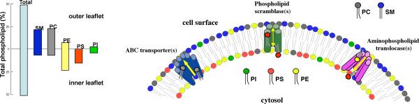
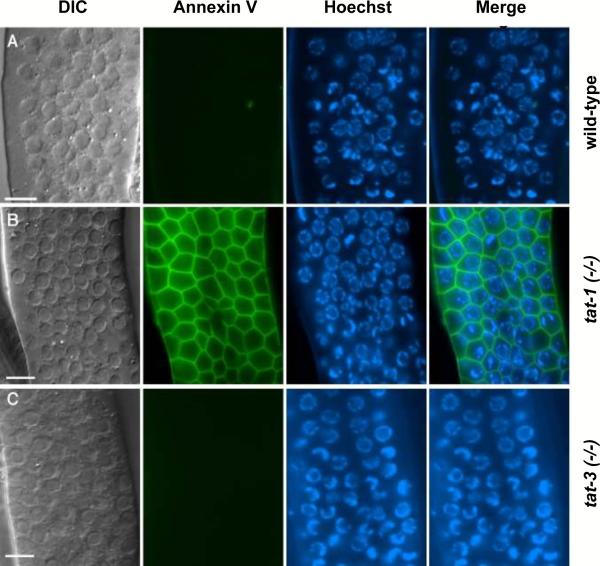
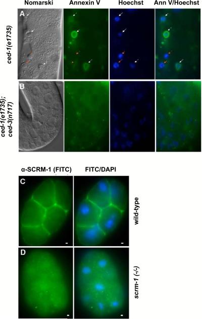
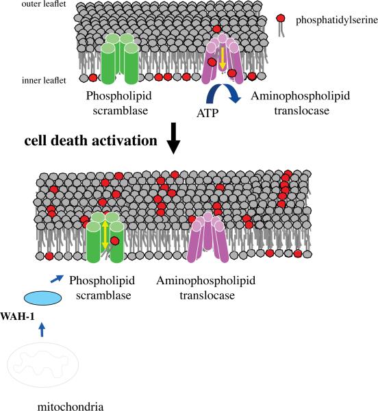
A common feature of all eukaryotic membranes is the non-random distribution of different lipid species in the lipid bilayer (lipid asymmetry). Lipid asymmetry provides the two sides of the plasma membrane with different biophysical properties and influences numerous cellular functions. Alteration of lipid asymmetry plays a prominent role during cell fusion, activation of the coagulation cascade, and recognition and removal of apoptotic cell corpses by macrophages (programmed cell clearance). Here we discuss the origin and maintenance of phospholipid asymmetry, based on recent studies in mammalian systems as well as in Caenhorhabditis elegans and other model organisms, along with emerging evidence for a conserved role of mitochondria in the loss of lipid asymmetry during apoptosis. The functional significance of lipid asymmetry and its disruption during health and disease is also discussed.


Figures

Figure 1
Figure 1
Phospholipid asymmetry and related lipid-translocating enzymes. In the plasma membrane of normal eukaryotic cells, phosphatidylcholine (PC) and sphingomyelin (SM) are present predominantly in the outer leaflet. Phosphatidylethanolamine (PE) and phosphatidylinositol (PI) reside mainly in the inner leaflet, while phosphatidylserine (PS) is located almost exclusively in the inner leaflet of the plasma membrane. Phospholipid asymmetry may be maintained or altered through the action of three classes of proteins, as discussed in the present review: phospholipid scramblases, ATP-binding cassette (ABC) transporters, and aminophospholipid translocases. The graph shows the transbilayer distribution of phospholipids across the human erythrocyte membrane (adapted from Daleke 2008).
Figure 2
Figure 2
Exposure of PS on the surface of C. elegans germ cells in tat-1-deficient worms. Exposed gonads of the following hermaphrodite adult worms were stained with the PS-binding protein, annexin V: (A) wild-type, (B) tat-1(tm1034), and (C) tat-3(tm1275), as described in Darland-Ransom et al. (2008). Images of differential contrast interference (DIC), annexin V staining, Hoechst 33342 staining, and the merged image of annexin V plus Hoechst 33342 staining are shown. The data show that reduction of tat-1 activity, but not of related tat-3 activity is sufficient to disrupt asymmetrical PS distribution on the surface of C. elegans germ cells. Scale bars correspond to 6.5 μm.
Figure 3
Figure 3
wah-1 and scrm-1 promote PS exposure on the surface of apoptotic germ cells in C. elegans. (A, B) The exposed gonad of a ced-1(e1735) (A) hermaphrodite animal or a ced-1(e1735); ced-3(n717) (B) animal was stained with annexin V and Hoechst 33342. Germ cell corpses were identified by their raised-button-like morphology under Nomarski optics and the condensed Hoechst 33342 staining pattern, as detailed in Wang et al. (2007). Germ cell corpses stained with both Hoechst and annexin V are indicated by white arrows, and those that were stained with Hoechst but not with annexin V are indicated with red arrows. The scale bar represents 6.5 μm. (C, D) SCRM-1 localizes to the plasma membrane. Images of FITC (SCRM-1 antibody staining) and FITC-DAPI C. elegans 16-cell stage wild-type (C) or scrm-1(tm805) (D) embryos are shown. The scale bars correspond to 1 μm.
Figure 4
Figure 4
Plasma membrane lipid transporters regulate the distribution of the phospholipid, PS. In quiescent cells, energy (ATP)-dependent translocation of PS from the outer to the inner leaflet of the plasma membrane serves to maintain the absolute asymmetry of PS distribution. Upon activation of the cell death program, the mitochondrial apoptogenic factor, WAH-1 (worm homolog of AIF) exits from mitochondria and activates the lipid scrambling activity of the plasma membrane phospholipid scramblase, SCRM-1. The bi-directional scrambling of phospholipids destroys the asymmetrical distribution of PS. Externalized PS serves as a signal to trigger engulfment by phagocytes through its binding to the PS receptor, PSR-1 (not shown).

References

    1. Arroyo A, Modriansky M, Serinkan FB, Bello RI, Matsura T, Jiang J, Tyurin VA, Tyurina YY, Fadeel B, Kagan VE. NADPH oxidase-dependent oxidation and externalization of phosphatidylserine during apoptosis in Me2SO-differentiated HL-60 cells. Role in phagocytic clearance. J Biol Chem. 2002;277:49965–49975. - PubMed
    1. Arur S, Uche UE, Rezaul K, Fong M, Scranton V, Cowan AE, Mohler W, Han DK. Annexin I is an endogenous ligand that mediates apoptotic cell engulfment. Dev Cell. 2003;4:587–598. - PubMed
    1. Asano K, Miwa M, Miwa K, Hanayama R, Nagase H, Nagata S, Tanaka M. Masking of phosphatidylserine inhibits apoptotic cell engulfment and induces autoantibody production in mice. J Exp Med. 2004;200:459–467. - PMC - PubMed
    1. Aussel C, Pelassy C, Breittmayer JP. CD95 (Fas/APO-1) induces an increased phosphatidylserine synthesis that precedes its externalization during programmed cell death. FEBS Lett. 1998;431:195–199. - PubMed
    1. Bassé F, Stout JG, Sims PJ, Wiedmer T. Isolation of an erythrocyte membrane protein that mediates Ca2+-dependent transbilayer movement of phospholipid. J Biol Chem. 1996;271:17205–17210. - PubMed

Publication types

LinkOut - more resources