Review
. 2009 Sep 28:11:e28.
doi: 10.1017/S1462399409001203.

Sarcoglycanopathies: molecular pathogenesis and therapeutic prospects

Affiliations
Free PMC article
Review

Sarcoglycanopathies: molecular pathogenesis and therapeutic prospects

Dorianna Sandonà et al. Expert Rev Mol Med. .
Free PMC article

Abstract

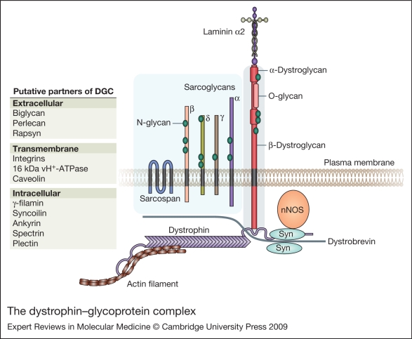
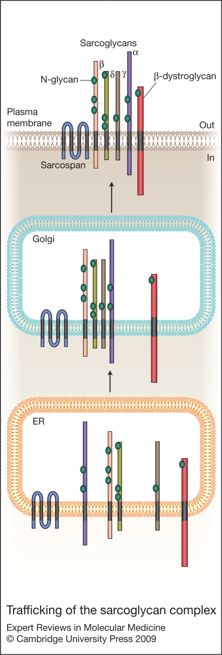
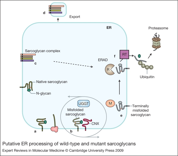
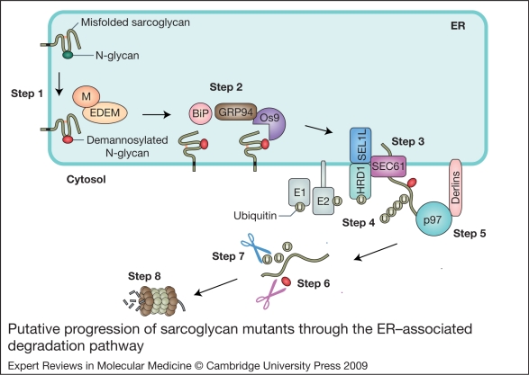
Sarcoglycanopathies are a group of autosomal recessive muscle-wasting disorders caused by genetic defects in one of four cell membrane glycoproteins, alpha-, beta-, gamma- or delta-sarcoglycan. These four sarcoglycans form a subcomplex that is closely linked to the major dystrophin-associated protein complex, which is essential for membrane integrity during muscle contraction and provides a scaffold for important signalling molecules. Proper assembly, trafficking and targeting of the sarcoglycan complex is of vital importance, and mutations that severely perturb tetramer formation and localisation result in sarcoglycanopathy. Gene defects in one sarcoglycan cause the absence or reduced concentration of the other subunits. Most genetic defects generate mutated proteins that are degraded through the cell's quality control system; however, in many cases, conformational modifications do not affect the function of the protein, yet it is recognised as misfolded and prematurely degraded. Recent evidence shows that misfolded sarcoglycans could be rescued to the cell membrane by assisting their maturation along the ER secretory pathway. This review summarises the etiopathogenesis of sarcoglycanopathies and highlights the quality control machinery as a potential pharmacological target for therapy of these genetic disorders.


Figures

Figure 1
Figure 1
The dystrophin–glycoprotein complex. Simplified scheme of dystrophin–glycoprotein complex (DGC) organisation. Dystrophin is localised in the cytoplasmic face of skeletal and cardiac cell membranes. Dystrophin binds actin filaments through two specific binding sites in its C-terminal domain and sites in the spectrin-like-repeat portion. The cysteine-rich domain assures the binding of dystrophin to the transmembrane β-dystroglycan, which in turn associates extracellularly with α-dystroglycan (blue box). α-dystroglycan interacts with laminin α2 and other cell matrix components, completing the backbone of the DGC. The sarcoglycan–sarcospan complex (light blue box), forms a lateral association with dystroglycans. N-glycan and O-glycan indicate, respectively, the N- and O-glycosylation moieties post-translationally added to sarcoglycans and dystroglycans. Additional sarcoglycan partners have been proposed (not all indicated), both intracellularly and extracellularly. Dystrobrevin, syntrophin (Syn) and neuronal nitric oxide synthase (nNOS) are intracellular components of the DGC. Many other proteins have been indicated to interact with DGC, either permanently or dynamically; a few of these are indicated. For a complete list of DGC components the reader should refer Refs , , , .
Figure 2
Figure 2
Trafficking of the sarcoglycan complex. The four sarcoglycans (α, β, γ and δ) are synthesised in the ER where they undergo cotranslational glycosylation (N-glycan) and proper conformational folding. Association of the four sarcoglycans occurs in the ER, with the βδ-sarcoglycan core being the trigger for the assembly of the tetrameric complex. The complex is then transported to the plasma membrane through the Golgi system, where it most likely assembles with sarcospan and dystroglycan. Once in the cell membrane, the other cytoplasmic components of the dystrophin–glycoprotein complex (DGC), aggregate to form the final structure.
Figure 3
Figure 3
Putative ER processing of wild-type and mutant sarcoglycans. Nascent sarcoglycans elongate into the ER where they undergo cotranslational glycosylation and proper conformational folding (a). Non-native polypeptides may experience repetitive rounds of folding within the calnexin (CNX)–calreticulin cycling system, a process regulated by UDP-glucose glycoprotein glucosyltransferase (UGGT) (b). A successfully folded protein then enters the maturation process, which comprises assembly with the other sarcoglycans to form a tetrameric complex (c). The protein complex is then exported from the ER (d). Misfolded proteins are degraded by endoplasmic reticulum-associated degradation (ERAD) by intervention of mannosidase I (M) (e). Individual components not able to assemble are also degraded (dotted arrow). ERAD occurs in a composite process of recognition (R), targeting for ER retrotranslocation (RT), and degradation of the terminally misfolded protein by the ubiquitin–proteasome system (f).
Figure 4
Figure 4
Putative progression of sarcoglycan mutants through the ER-associated degradation pathway. The putative processing of sarcoglycan mutants is represented in consecutive steps. Non-native sarcoglycans are targeted to ERAD by the intervention of mannosidase I and/or mannosidase-like proteins (EDEM), which remove a mannose from the N-glycan and terminate its maturation process (Step 1). Then, the misfolded sarcoglycan is identified and escorted for retrotranslocation by a targeting complex composed of three probable components (BiP, GRP94 and Os9) (Step 2). Dislocation of the ERAD substrate occurs through the SEL1L–HRD1 complex (Step 3), which also includes Sec61, the actual channel. During retrotranslocation, the polypeptide is ubiquitinated by cytosolic and membrane-associated E1, E2 and E3 enzymes (Step 4). Note that HRD1 is one such E3 ligase. A multimeric protein complex, which comprises the p97 ATPase and Derlin proteins (through which the complex attaches to the ER membrane), dislocates the terminally misfolded sarcoglycan from the ER (Step 5). The same p97 complex targets the polyubiquitinated (U) sarcoglycan to the 26S proteasome. Approaching the 19S cap of the proteasome, the N-glycan and the ubiquitin chain are removed by N-glycanase (Step 6) and deubiquitinating enzymes (Step 7). The polypeptide is eventually threaded into the 20S proteasome catalytic core where it is broken down into small fragments (Step 8).
Figure 5
Figure 5
Sarcoglycan missense mutations. Sequence of α-, β-, γ and δ-sarcoglycan with all missense substitutions responsible for LGMD indicated in red. The indicated missense mutations are derived from the Leiden database (http://www.lovd.org) and recent reports (Refs 53, 92). The stretch of residues of the transmembrane domain is shown in yellow, the putative N-linked glycosylation sites in green, and the predicted phosphorylation sites (Ref. 95) in dark green. In α-sarcoglycan, the signal sequence is indicated in italics, the cadherin-like domain (Ref. 76) in grey, whereas the putative ATP-binding site (Ref. 38) is shown in blue. In β-, γ- and δ-sarcoglycan, the putative EGF-like sequence (Ref. 3) is underlined.

Similar articles

Cited by

References

    1. Blake D.J.. et al. Function and genetics of dystrophin and dystrophin-related proteins in muscle. Physiological Reviews. 2002;82:291–329. - PubMed
    1. Davies K.E., Nowak K.J.. Molecular mechanisms of muscular dystrophies: old and new players. Nature Reviews. Molecular Cell Biology. 2006;7:762–773. - PubMed
    1. Hack A.A.. et al. Sarcoglycans in muscular dystrophy. Microscopy Research and Technique. 2000;48:167–180. - PubMed
    1. Ozawa E.. et al. Molecular and cell biology of the sarcoglycan complex. Muscle and Nerve. 2005;32:563–576. - PubMed
    1. Bönnemann C.G.. et al. β-Sarcoglycan (A3b) mutations cause autosomal recessive muscular dystrophy with loss of the sarcoglycan complex. Nature Genetics. 1995;11:266–273. - PubMed

Further reading, resources and contacts

Publications
    1. Straub V., Bushby K.. Therapeutic possibilities in the autosomal recessive limb-girdle muscular dystrophies. Neurotherapeutics. 2008;5:619–626. - PMC - PubMed
    2. This review describes an overview of potential novel therapeutical interventions in autosomal recessive limb-girdle muscular dystrophies.

    1. Muir L.A., Chamberlain J.S.. Emerging strategies for cell and gene therapy of the muscular dystrophies. Expert Reviews in Molecular Medicine. 2009;11:e8. - PMC - PubMed
    2. This review describes novel therapeutical interventions developed to cure Duchenne muscular dystrophy and that can be extended to other dystrophies.

    1. Linde L., Kerem B.. Introducing sense into nonsense in treatments of human genetic diseases. Trends in Genetics. 2008;24:552–563. - PubMed
    2. This review deals with therapeutical approaches aimed at curing nonsense mutations creating a premature stop codon.

    1. Braun S.. Muscular gene transfer using nonviral vectors. Current Gene Therapy. 2008;8:391–405. - PubMed
    2. This review reports on recent progresses of plasmid delivery of genes to muscles by using non-viral vectors.

Websites
    1. http://www.dmd.nl/sgca_seqvar.html http://www.dmd.nl/sgca_seqvar.html
    2. The Muscular Dystrophy Association, MDA, together with numerous international associates, provides information on muscular dystrophies and helpful resources for patients:

    1. http://www.mda.org/ http://www.mda.org/
    2. Treat Neuromuscular Disorders (TREAT-NMD) provide ample information about muscular dystrophies and updates on new therapeutic developments:

    1. http://www.treat-nmd.eu/home.php http://www.treat-nmd.eu/home.php
    2. The Center for Neuromuscular Diseases, CNMD, is dedicated to clinical trials and therapy:

    1. http://www.cnmd.ac.uk/index.html http://www.cnmd.ac.uk/index.html
    2. ClinicalTrialsGov offers a complete list of worldwide clinical trials on muscular dystrophies, and other medical conditions:

    1. http://clinicaltrials.gov/ct2/home http://clinicaltrials.gov/ct2/home

Publication types

MeSH terms

Substances