. 2009 Dec;1793(12):1886-93.
doi: 10.1016/j.bbamcr.2009.09.013. Epub 2009 Sep 23.

Calcium-dependent plasma membrane repair requires m- or mu-calpain, but not calpain-3, the proteasome, or caspases

Affiliations

Calcium-dependent plasma membrane repair requires m- or mu-calpain, but not calpain-3, the proteasome, or caspases

Ronald L Mellgren et al. Biochim Biophys Acta. 2009 Dec.

Abstract

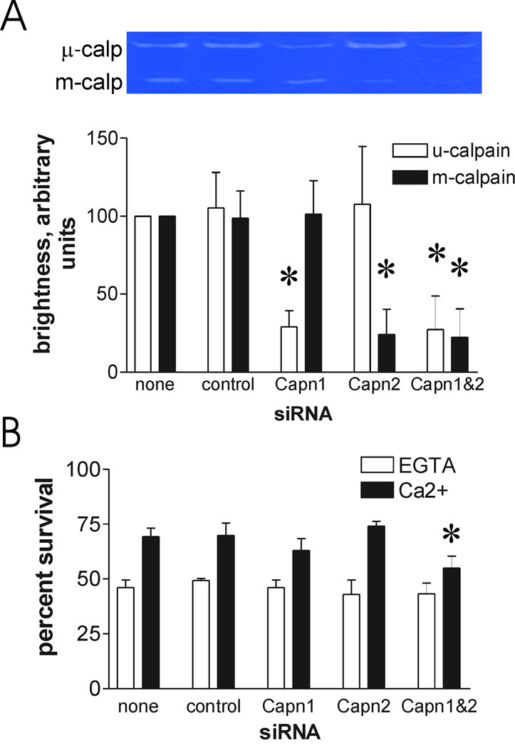
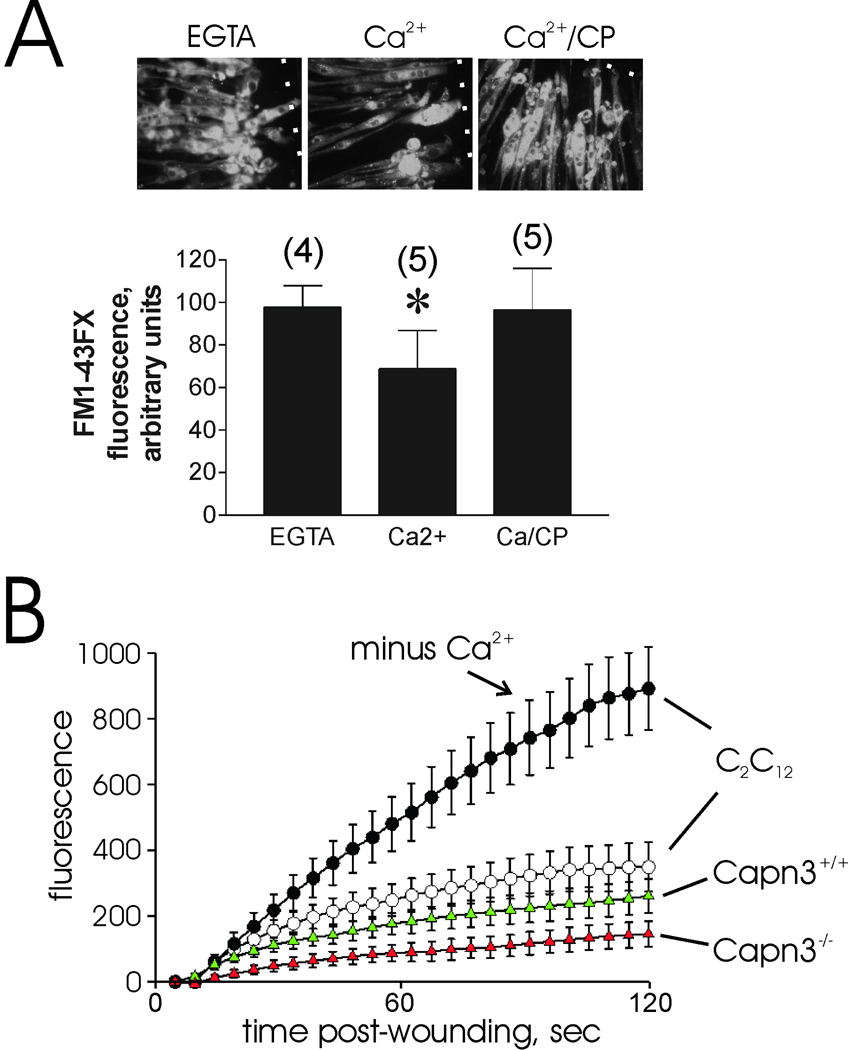
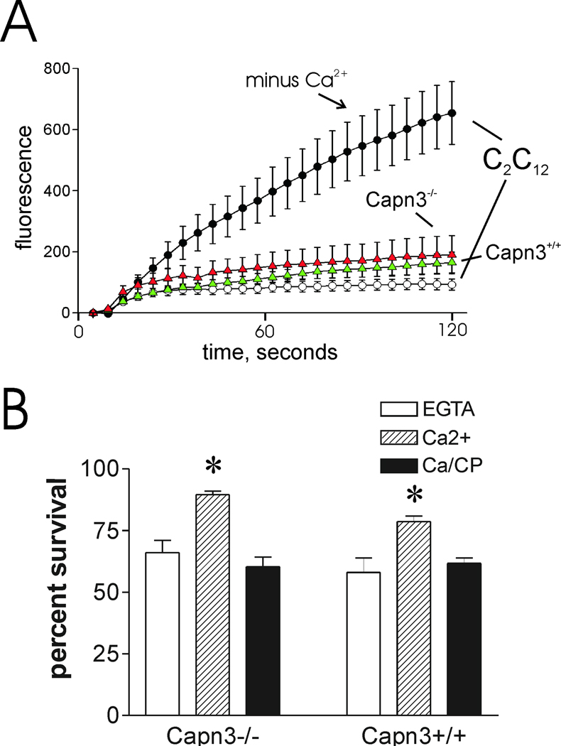
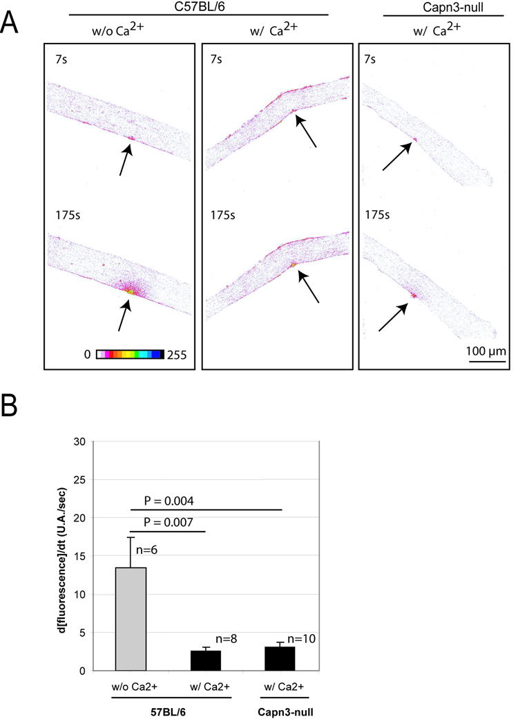
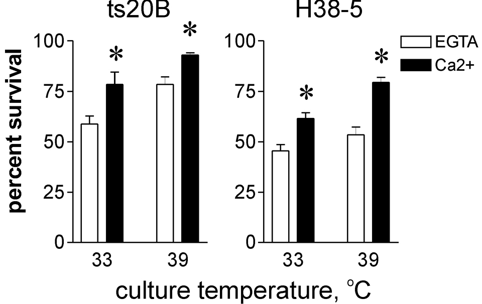
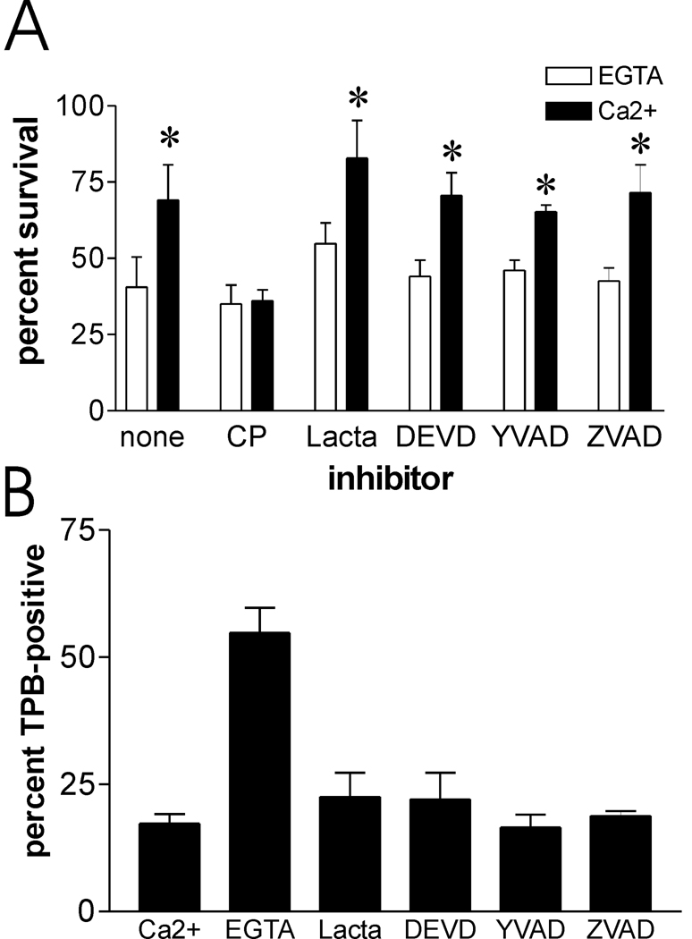
Mechanically damaged plasma membrane undergoes rapid calcium-dependent resealing that appears to depend, at least in part, on calpain-mediated cortical cytoskeletal remodeling. Cells null for Capns1, the non-catalytic small subunit present in both m- and mu-calpains, do not undergo calcium-mediated resealing. However, it is not known which of these calpains is needed for repair, or whether other major cytosolic proteinases may participate. Utilizing isozyme-selective siRNAs to decrease expression of Capn1 or Capn2, catalytic subunits of mu- and m-calpains, respectively, in a mouse embryonic fibroblast cell line, we now show that substantial loss of both activities is required to compromise calcium-mediated survival after cell scrape-damage. Using skeletal myotubes derived from Capn3-null mice, we were unable to demonstrate loss of sarcolemma resealing after needle scratch or laser damage. Isolated muscle fibers from Capn3 knockout mice also efficiently repaired laser damage. Employing either a cell line expressing a temperature sensitive E1 ubiquitin ligase, or lactacystin, a specific proteasome inhibitor, it was not possible to demonstrate an effect of the proteasome on calcium-mediated survival after injury. Moreover, several cell-permeant caspase inhibitors were incapable of significantly decreasing survival or inhibiting membrane repair. Taken together with previous studies, the results show that m- or mu-calpain can facilitate repair of damaged plasma membrane. While there was no evidence for the involvement of calpain-3, the proteasome or caspases in early events of plasma membrane repair, our studies do not rule out their participation in downstream events that may link plasma membrane repair to adaptive remodeling after injury.


Figures

Figure 1
Figure 1
The Capns1−/−R rescued fibroblast cell line expresses limited calpain activity, but shows partial recovery of calcium-mediated cell survival after scraping. A. Casein zymogram of Capns1 cell lines. Cell lysates were subjected to zymography as described in the Materials and Methods section. The μ- and m-calpain bands are shown, as assessed by the migration of purified human μ- and bovine m-calpains, respectively. Lanes are from the same zymogram, but were separated to prevent sample “bleedover”. B. Relative intensity of m-calpain bands from three independent zymography experiments, using the NIH imageJ software program to estimate brightness of the cleared casein zones. C. Capns1−/−R cells were scraped with the indicated additions, as described in the Materials and Methods section, and allowed to recover for 12 hours before assessing cell survival. CP=20 µM calpeptin. Asterisk: P = 0.002 vs EGTA sample (N=6). D. Capns1−/−, Capns1−/−R, and Capns1+/+ fibroblasts were cultured to 30–50% confluence, and scraped in the presence of calcium. Asterisk: P<0.001 vs Capns1−/−. Double asterisk: P<0.001 vs Capns1−/−R.
Figure 2
Figure 2
Calcium-dependent survival after scraping requires the typical, ubiquitous calpains. A. Capns1+/+ fibroblasts were transfected with the indicated siRNAs. After 72 hours, the cells were lysed and subjected to casein zymography to assess loss of μ- and m-calpain activity. The inset shows results of one experiment. The graph presents data from three experiments. Asterisks: P<0.02 vs corresponding control siRNA. B. Other samples were scraped and viability was determined 16 hours later. Details are provided in the Materials and Methods section. Asterisk: P = 0.005 vs no siRNA addition (N=4).
Figure 3
Figure 3
Calcium-dependent repair of damaged sarcolemma is not compromised in myotubes derived from Capn3-null mice. A. Myotubes prepared from Capn3−/− myoblasts were subjected to needle scratch damage in the presence of FM1-43FX dye. The dotted white lines indicate the sites of scratch injury. Dye uptake for the first minute after damage was semi-quantitatively assessed as described in the Materials and Methods section (asterisk: P = 0.014 vs EGTA samples). Numbers in parentheses refer to the total number of scratch sites analyzed in two different culture dish wells. Upper panel depicts typical results for each injury condition. B. Laser injury of Capn3−/− and Capn3+/+ myotubes, as described in the Materials and Methods section. Except where noted, cells were injured in the presence of 1 mM CaCl2. As noted in many previous studies, calcium-mediated resealing of wounded membrane rapidly prevents accumulation of dye, as shown by injury of C2C12 myotubes in the presence of calcium compared with EGTA. Note that both Capn3−/− and Capn3+/+ myotubes (red and green triangles, respectively) resealed efficiently.
Figure 4
Figure 4
Membrane repair and calcium-dependent cell survival are not significantly compromised in injured Capn3−/− myoblasts. Myoblasts were subjected to laser injury (A) or scrape injury (B) using the standard protocols for each (see Materials and Methods section). Symbols are the same as in figure 3. Asterisks: P < 0.005 vs EGTA samples.
Figure 5
Figure 5
Comparison of membrane repair capacity between Capn3-null and C57BL/6 mice A. A membrane repair assay was performed on single muscle fibers from C57BL/6 and Capn3-null mice. Images are represented in a “16 colors inverted” look up table (table depicted at the bottom of the left panel) from ImageJ software. Arrows indicate the site of injury. The time at which each picture was taken is also indicated. The two left panels show a representative assay performed on C57BL/6 fibers without (w/o) or with (w/) Ca2+. The right panel shows a representative repair assay performed on Capn3-null fibers. Scale bar = 100 µm. B. Histogram representing the rates of fluorescence influx (Δ[fluorescence]/Δt) in fibers of C57BL/6 w/o Ca2+, w/ Ca2+ and Capn3-null w/ Ca2+. The fibers were sampled from 3 different mice for each condition. Data are means ± s.e.m. Statistical analysis was performed with Student's t-test.
Figure 6
Figure 6
Recovery after scrape-damage is not compromised in a ubiquitin ligase mutant cell line. A ubiquitin E1 ligase temperature sensitive mutant fibroblast cell line (ts20B) and H38-5 control cell line were cultured at permissive (33 °C) or restrictive (39 °C) temperatures prior to scrape-damage and survival analysis as described in the Materials and Methods section. Asterisks: P ≤ 0.02 vs paired EGTA samples (N=4).
Figure 7
Figure 7
Proteasome and caspases do not appear to contribute to survival after scrape damage, or to acute repair of damaged plasma membrane. A. Capns1+/+ cells were preincubated with protease inhibitors as described in the Materials and Methods section, and then subjected to scrape damage in the presence of calcium and inhibitors. Concentrations of inhibitor used were: calpeptin (CP), 20 µM; lactacystin (lacta), 5 µM; DEVD, 2 µM; YVAD, 2 µM; ZVAD, 10µM. Asterisks: P < 0.01 vs paired EGTA samples (N=4). B. Capns1+/+ cells were treated with inhibitors, scraped in the presence of calcium, and analyzed for trypan blue (TPB) uptake one minute after scraping.

References

    1. McNeil PL, Steinhardt RA. Plasma membrane disruption: repair, prevention, adaptation. Annu Rev Cell Dev Biol. 2003;19:697–731. - PubMed
    1. McNeil PL, Kirchhausen T. An emergency response team for membrane repair. Nat Rev Mol Cell Biol. 2005;6:499–505. - PubMed
    1. Han R, Bansal D, Miyake K, Muniz VP, Weiss RM, McNeil PL, Campbell KP. Dysferlin-mediated membrane repair protects the heart from stress-induced left ventricular injury. J Clin Invest. 2007;117:1805–1813. - PMC - PubMed
    1. McNeil PL, Khakee R. Disruptions of muscle fiber plasma membranes. Role in exercise-induced damage. Am J Pathol. 1992;140:1097–1109. - PMC - PubMed
    1. Fischer TA, McNeil PL, Khakee R, Finn P, Kelly RA, Pfeffer MA, Pfeffer JM. Cardiac myocyte membrane wounding in the abruptly pressure-overloaded rat heart under high wall stress. Hypertension. 1997;30:1041–1046. - PubMed

Publication types