. 2010 Jan;24(1):153-9.
doi: 10.1016/j.bbi.2009.09.011. Epub 2009 Sep 23.

Repeated bouts of aerobic exercise enhance regulatory T cell responses in a murine asthma model

Affiliations

Repeated bouts of aerobic exercise enhance regulatory T cell responses in a murine asthma model

Thomas Lowder et al. Brain Behav Immun. 2010 Jan.

Abstract

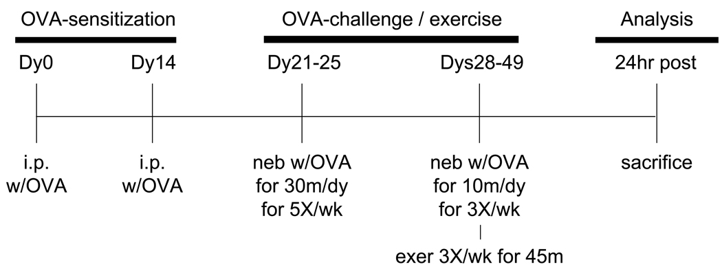
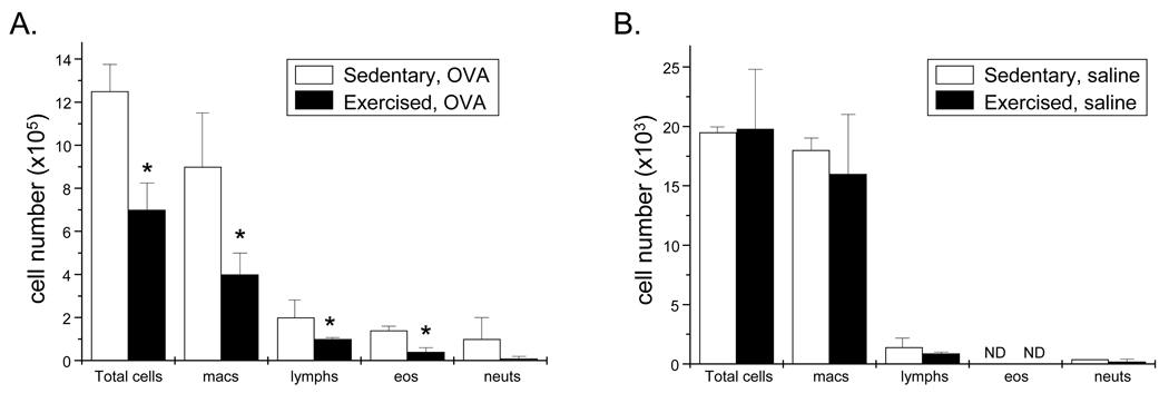
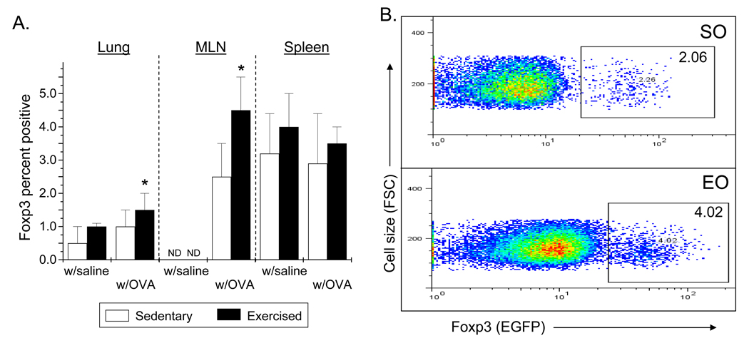
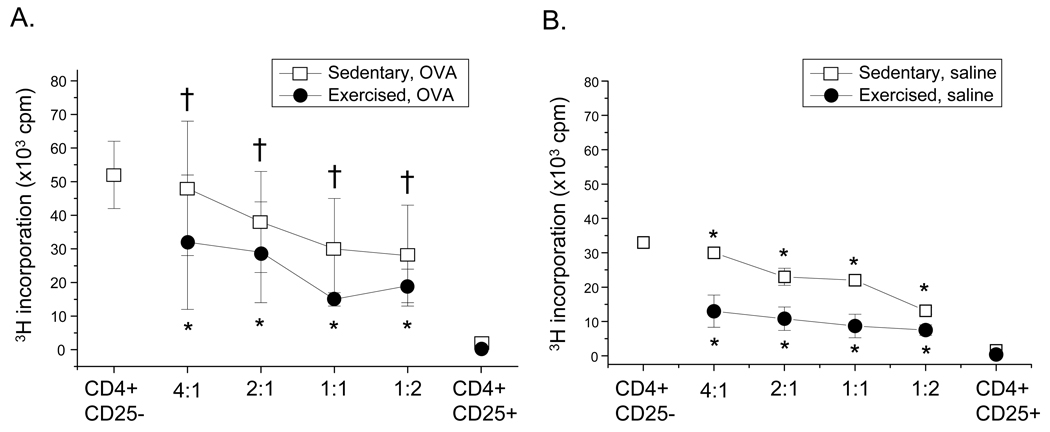
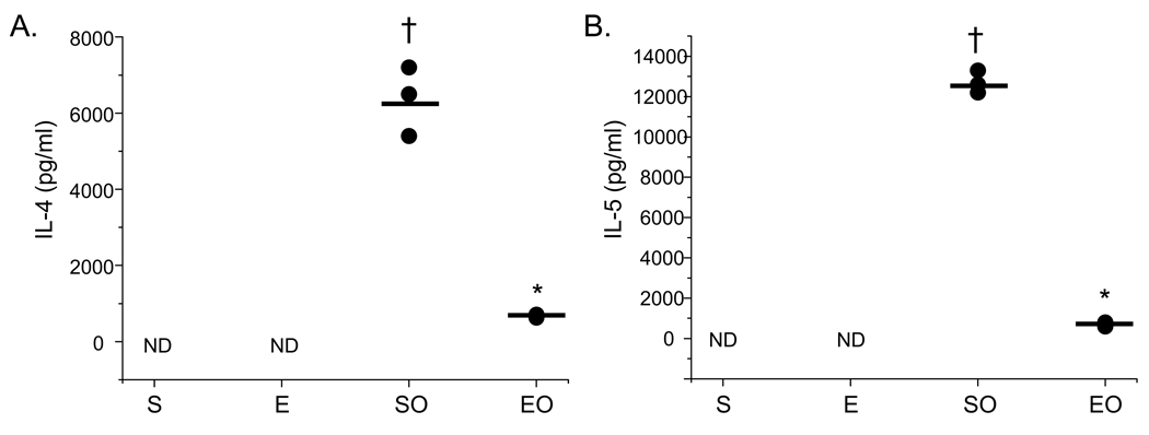
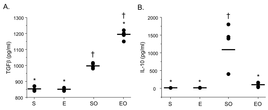
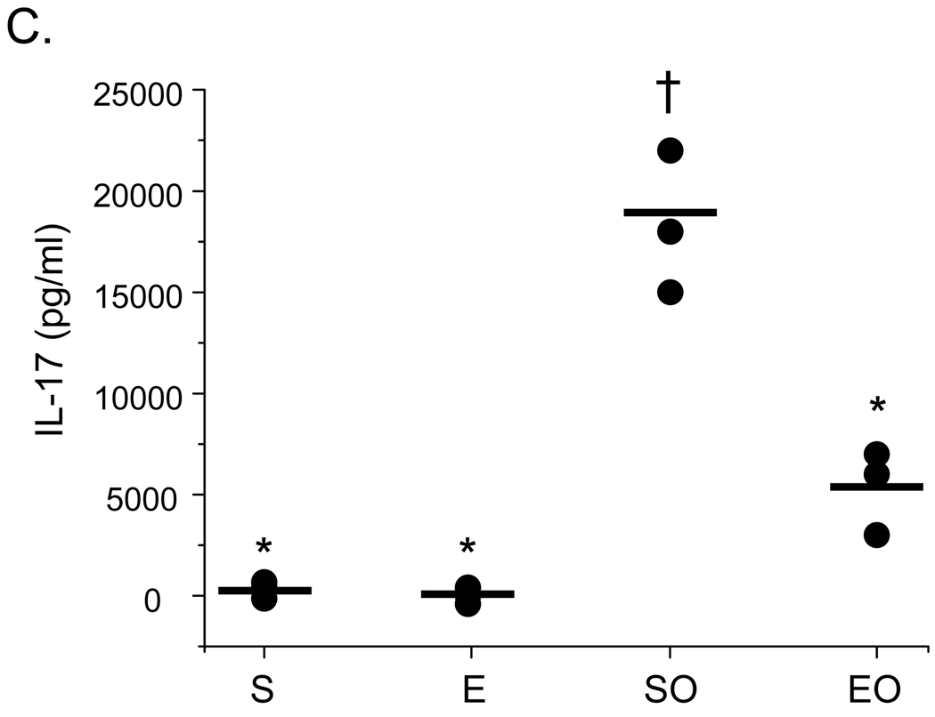
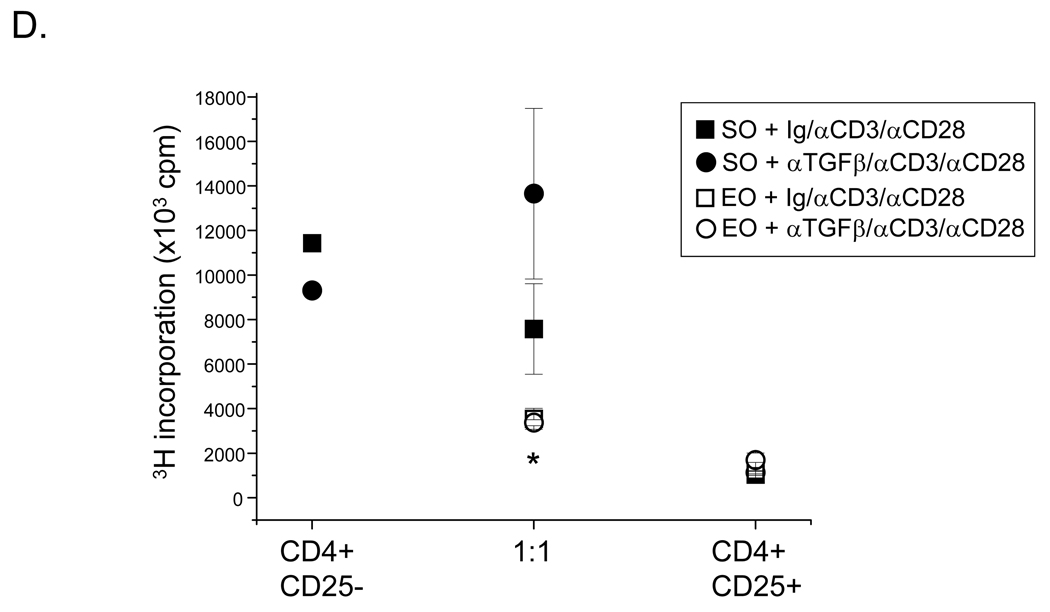
We have reported previously that moderate intensity aerobic exercise training attenuates airway inflammation in a murine asthma model. Recent studies implicate regulatory T (Treg) cells in decreasing asthma-related airway inflammation; as such, the current study examined the effect of exercise on Treg cell function in a murine asthma model. Mice were sensitized with ovalbumin (OVA) prior to the start of exercise training at a moderate intensity 3x/week for 4weeks; exercise was performed as treadmill running (13.5m/min, 0% grade). Mice were OVA challenged repeatedly throughout the exercise protocol. At protocol completion, mice were analyzed for changes in the number and suppressive function of CD4(+)CD25(+)Foxp3(+) cells isolated from lungs, mediastinal lymph nodes, and spleens. Results show that exercise increased significantly the number of Foxp3(+) cells within the lungs and mediastinal lymph nodes, but not the spleens, of OVA-treated mice as compared with sedentary controls. Exercise also enhanced the suppression function of CD4(+)CD25(+)Foxp3(+) Treg cells derived from OVA-treated mice as compared with sedentary controls. Specifically, Treg cells from exercised, OVA-treated mice more effectively suppressed CD4(+)CD25(-) cell proliferation and Th2 cytokine production in vitro. Enhanced suppression was associated with increased protein levels of TGF-beta and lesser amounts of IL-10 and IL-17; however, blocking TGF-beta had no effect on suppressive functions. These data demonstrate that exercise-mediated increases in Treg cell function may play a role in the attenuation of airway inflammation. Further, these results indicate that moderate intensity aerobic exercise training may alter the Treg cell function within the asthmatic airway.


Conflict of interest statement

Conflict of interest statement: All authors declare that there are no conflicts of interest

Figures

Figure 1
Figure 1
Time-line of OVA treatment and exercise training. As described in the Methods section, mice were injected with OVA twice on dys 0 and 14 followed OVA aerosolizations on dys 21 – 25. Mice were then challenged repeatedly with OVA via aerosolization during exercise training on dys 28 – 49. 24hr post the last OVA/exercise bout, mice were sacrificed and analyzed as indicated.
Figure 2
Figure 2
Exercise decreases cellular infiltration in the lungs of OVA-treated, Foxp3 reporter mice. 24 hr post protocol completion, BAL was performed and resulting lavagates were analyzed for changes in total number of cells, macrophages, lymphocytes, eosinophils, and neutrophils. Results are reported as A) cell number × 105 (OVA-treated) and B) cell number × 103 (saline-treated) (*p < 0.03 as compared with OVA-treated, sedentary mice via unpaired Student’s T-test; n = 3 – 6 mice per group; ND – none detected). Two-way ANOVA indicated that OVA-treatment significantly increased cell numbers in all groups (p = 0.001, F = 26). OVA groups differed from saline groups as determined via Tukey’s post hoc analysis (p < 0.05).
Figure 3
Figure 3
Exercise increases the number of CD4+Foxp3+ cells in the lungs and mediastinal lymph nodes (MLNs) of OVA-treated mice. Foxp3+ reporter mice were OVA-treated and exercised as described in Methods. 24hr post protocol completion, cells were isolated from lungs, MLN, and spleens and analyzed via flow cytometry. A) Results are presented as percent Foxp3+ cells (*p = 0.01 as compared with SO mice via unpaired Student’s T-test; n = 3 – 14 per group; ND – none detected). Two-way ANOVA analysis indicated that exercise significantly increased the number of Foxp3+ cells in the in lung and spleen (p ≤ 0.02, F ≥ 5). For the lung and spleen samples, exercise groups differed from sedentary groups as determined via Tukey’s post hoc (p < 0.05). B) Representative histograms from the analyses of MLN (SO – sedentary, OVA-treated; EO – exercised, OVA-treated).
Figure 4
Figure 4
Exercise increases the suppressive function of CD4+CD25+Foxp3+ cells. Foxp3+ reporter mice were OVA-treated and exercised as described in Methods. 24 hr post-protocol completion, CD4+CD25+Foxp3+ cells were isolated from A) spleens of OVA-treated mice (n = 16 – 22 mice per group) as well as B) spleens of saline-treated mice (n = 3 – 4 samples per group) via MACS separation. Cells were co-cultured at increasing concentrations (effector:suppressor) with CD4+CD25 cells derived from sedentary, OVA-treated (SO) mice in the presence of anti-CD3 and anti-CD28 for 72 hrs. Co-cultures were pulsed for an additional 18h with [3H]thymidine and analyzed for suppression. Results are presented as 3H incorporation cpm. Two-way ANOVA analysis indicated that increased suppressive function is the result of exercise and not OVA treatment or cell ratio (exercise p<0.0001; F ≥ 10). An unpaired Student’s T test showed *p < 0.01 as compared with SO mice and †p < 0.01 as compared with S mice. Exercise groups differed from sedentary groups as determined via Tukey’s post hoc analysis (p < 0.0001).
Figure 5
Figure 5
Exercise decreases the protein levels of IL-4 and IL5 in CD4+CD25+Foxp3+ Treg cell suppression cultures. Supernatants were harvested from parallel cultures described in Figure 4 and analyzed for A) IL-4 and B) IL-5 protein content via ELISA (S – sedentary; E – exercised; SO – sedentary, OVA-treated; EO – exercised, OVA-treated). Results are presented as pg/ml (*p < 0.04 as compared with SO mice, †p < 0.04 as compared with S mice as determined via unpaired Student’s T-test; n = 3 – 6 samples per group; ND – none detected).
Figure 6
Figure 6
Exercise increases the protein levels of TGF-β, but not IL-10 or IL-17, in CD4+CD25+Foxp3+ Treg cell suppression cultures. Supernatants were harvested from parallel cultures described in Figure 4 and analyzed for A) TGF-β, B) IL-10, and C) IL-17 protein content via ELISA or multiplex assay (S – sedentary; E – exercised; SO – sedentary, OVA-treated; EO – exercised, OVA-treated). Results are presented as pg/ml (*p < 0.05 as compared with SO mice, †p < 0.05 as compared with S mice determined via unpaired Student’s T-test; n = 3 – 6 samples per group; each sample contained cells pooled from 3 – 4 mice). For TGFβ, two-way ANOVA analysis revealed significance of exercise at p = 0.03 and significance of OVA at p = 0.0001 (F = 69). Exercise groups differed from sedentary groups as determined via Tukey’s post hoc analysis (p ≤ 0.05). D) For anti-TGFβ blocking studies, an anti-TGFβ blocking antibody or isotype-matched antibody control was added to suppression assays, and analyzed as described in Figure 4. Results are presented as 3H incorporation (cpm; *p < 0.01 as compared with SO mice determined via unpaired Student’s T-test; n = 3 – 5 samples per group).
Figure 6
Figure 6
Exercise increases the protein levels of TGF-β, but not IL-10 or IL-17, in CD4+CD25+Foxp3+ Treg cell suppression cultures. Supernatants were harvested from parallel cultures described in Figure 4 and analyzed for A) TGF-β, B) IL-10, and C) IL-17 protein content via ELISA or multiplex assay (S – sedentary; E – exercised; SO – sedentary, OVA-treated; EO – exercised, OVA-treated). Results are presented as pg/ml (*p < 0.05 as compared with SO mice, †p < 0.05 as compared with S mice determined via unpaired Student’s T-test; n = 3 – 6 samples per group; each sample contained cells pooled from 3 – 4 mice). For TGFβ, two-way ANOVA analysis revealed significance of exercise at p = 0.03 and significance of OVA at p = 0.0001 (F = 69). Exercise groups differed from sedentary groups as determined via Tukey’s post hoc analysis (p ≤ 0.05). D) For anti-TGFβ blocking studies, an anti-TGFβ blocking antibody or isotype-matched antibody control was added to suppression assays, and analyzed as described in Figure 4. Results are presented as 3H incorporation (cpm; *p < 0.01 as compared with SO mice determined via unpaired Student’s T-test; n = 3 – 5 samples per group).

References

    1. Beriou G, Costantino C, Ashley C, Yang L, Kuchroo V, Baecher-Allan C, et al. IL-17 producing human peripheral regulatory T cells retain suppressive function. Blood. 2009 (in press) - PMC - PubMed
    1. Chen X, Oppenheim J, Winkler-Pickett R, Ortaldo J, Howard O. Glucocorticoid amplifies IL-2-dependent expansion of functional FoxP3+CD4+CD25+ T regulatory cells in vivo and enhances their capacity to suppress EAE. Eur J Immunol. 2006;36:2139–2149. - PubMed
    1. Fontenot J, Rudensky A. A well adapted regulatory contrivance: regulatory T cell development and the foxhead family transciption factor Foxp3. Nat.Immunol. 2005;6:331–337. - PubMed
    1. Heijink I, Van Oosterhout A. Strategies for targeting T-cells in allergic disease and asthma. Pharmacol Ther. 2006;112:489–500. - PubMed
    1. Hewitt M, Creel A, Estell K, Davis I, Schwiebert L. Acute exercise decreases airway inflammation, but not responsiveness, in an allergic asthma model. Am J Respir Cell Mol Biol. 2009;40:83–89. - PMC - PubMed

Publication types

MeSH terms