Case Reports
. 2009 Oct;85(4):521-7.
doi: 10.1016/j.ajhg.2009.09.001. Epub 2009 Sep 24.

PPIB mutations cause severe osteogenesis imperfecta

Affiliations
Case Reports

PPIB mutations cause severe osteogenesis imperfecta

Fleur S van Dijk et al. Am J Hum Genet. 2009 Oct.

Abstract

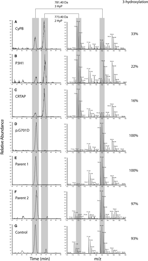
Deficiency of cartilage-associated protein (CRTAP) or prolyl 3-hydroxylase 1(P3H1) has been reported in autosomal-recessive lethal or severe osteogenesis imperfecta (OI). CRTAP, P3H1, and cyclophilin B (CyPB) form an intracellular collagen-modifying complex that 3-hydroxylates proline at position 986 (P986) in the alpha1 chains of collagen type I. This 3-prolyl hydroxylation is decreased in patients with CRTAP and P3H1 deficiency. It was suspected that mutations in the PPIB gene encoding CyPB would also cause OI with decreased collagen 3-prolyl hydroxylation. To our knowledge we present the first two families with recessive OI caused by PPIB gene mutations. The clinical phenotype is compatible with OI Sillence type II-B/III as seen with COL1A1/2, CRTAP, and LEPRE1 mutations. The percentage of 3-hydroxylated P986 residues in patients with PPIB mutations is decreased in comparison to normal, but it is higher than in patients with CRTAP and LEPRE1 mutations. This result and the fact that CyPB is demonstrable independent of CRTAP and P3H1, along with reported decreased 3-prolyl hydroxylation due to deficiency of CRTAP lacking the catalytic hydroxylation domain and the known function of CyPB as a cis-trans isomerase, suggest that recessive OI is caused by a dysfunctional P3H1/CRTAP/CyPB complex rather than by the lack of 3-prolyl hydroxylation of a single proline residue in the alpha1 chains of collagen type I.


Figures

Figure 1
Figure 1
Radiological Features of Affected Probands from Families 1 and 2 (A) Fetal anteroposterior radiographs of proband 1 from family 1 at 21 + 2 weeks of gestation show the following: normal skull mineralization for gestational age; slender ribs without fractures; incomplete ossification of T5 and T12; somewhat irregular proximal metaphyses of the humeri, radii, and ulnae; and bowing of the ulnae. Bowed femora with fractures and some loss of modeling and bowed tibiae and fibula, possibly with fractures, are apparent. The radiological features are compatible with Sillence OI type II-B/III. (B and C) Radiographs of proband 2 from family 2 show normal mineralization of the skull, fractures of both humeri and radii and the left ulna with callus formation and some loss of modeling of the humeri. Discontinuously beaded ribs and a small, bell-shaped thorax are noted. Multiple fractures with callus formation of femora, tibiae, and fibula exist with moderate loss of modeling of the femora. The radiological features are compatible with Sillence OI type II-B/III.
Figure 2
Figure 2
PPIB Mutations in Severe OI (A) A homozygous c.556–559 delAAGA in exon 5 resulting in p.Lys186GlnfsX8 is shown in probands of family 1. (B) A homozygous c.451 C > T mutation in exon 4 resulting in p.Gln151X is shown in probands of family 2.
Figure 3
Figure 3
Immunoblot in Family 1 An immunoblot is shown of fibroblast CyPB, which is lacking in the affected infant P1-1 and present in the parent and the control (polyclonal antibodies raised against the whole protein were used in this immunoblot, 11607-1-AP, Proteintech, Manchester, UK). CRTAP is present in P1-1, the parent, and the control.
Figure 4
Figure 4
Results of Immunohistochemistry with Antibodies against CRTAP, P3H1, and CyPB Immunohistochemistry performed on bone tissue with CyPB-CRTAP and P3H1 antibodies shows the following: (A–C) a positive signal of CyPB, CRTAP, and P3H1 in a control without OI; (D–F) a positive signal of CyPB, CRTAP, and P3H1 in a patient with a COL1A1 mutation (c.1238G > A [p.Gly413Asp]); (G–I) no signal of either CRTAP or P3H1 but a signal of CyPB in a patient with a homozygous c.404 delG CRTAP mutation; (J–L) no signal of either P3H1 or CRTAP and a signal of CyPB in a patient with a homozygous c.401_402 delAA LEPRE1 mutation; and (M–O) no signal of CyPB but signals of both CRTAP and P3H1 antibodies in P1-2 with a homozygous PPIB mutation. AB: antibody.
Figure 5
Figure 5
Collagen I α-1 P986 3-Hydroxylation Level in Patients with OI Due to Different Molecular Genetic Causes Figure 5 shows mass spectrometry analysis of the C1A1 P986-containing tryptic peptide, modified with either two or three hydroxyl groups (as indicated by an asterisk below the modified proline residue in Figure S2) in patients with homozygous PPIB (A), LEPRE1 (B), or CRTAP (C) mutations or a heterozygous COL1A1 mutation (D), in the parents of the patient with PPIB mutations (E and F), and in a control (G). The left panel shows extracted ion chromatograms (XIC), generated at m/z 773.4 and 781.4 ± 0.05, and the right panel shows the corresponding summed mass spectra (40–50 min.). XIC peak areas were determined for doubly and triply hydroxylated peptides. The C1A1 P986 3-hydroxylation level (%) was calculated as triple-hydroxylation area (triple-hydroxylation area + double-hydroxylation area). Collagen I α-1 peptides that show normal P986 3-hydroxylation have an m/z ratio of 781.4, whereas peptides lacking 3-hydroxylation have an m/z ratio of 773.4. Controls (D–F) show normal 3-hydroxylation (>90%). P1-1 with homozygous PPIB mutations (A) shows decreased 3-hydroxylation (33%). Patients with homozygous LEPRE1 (p.Glu134fs) or CRTAP (p.Ala8fs) mutations (B and C, respectively), however, show even more decreased 3-hydroxylation levels (22% and 16%, respectively).
Figure 6
Figure 6
Electrophoresis of Collagen Type I Chains in Fibroblasts and Chorionic Villi of Patients with a Homozygous CRTAP, LEPRE1, or PPIB Mutation or a Heterozygous COL1A1 Mutation Backstreaking and/or increased band width is visible in cells (c) and media (m), indicating overmodification of collagen type I chains in chorionic villi cells and fibroblasts of patients with lethal or severe OI due to homozygous CRTAP (p.Ala8fs), LEPRE1 (p.Glu134fs), or PPIB (p.Lys186GlnfsX8) mutations or a heterozygous COL1A1 (p.G965S in chorionic villi cells and p.G662V in fibroblasts) mutation. The slight apparent overmodification of other collagens appears to be an effect typically evident in chorionic villi.

References

    1. Marini J.C., Cabral W.A., Barnes A.M., Chang W. Components of the collagen prolyl 3-hydroxylation complex are crucial for normal bone development. Cell Cycle. 2007;6:1675–1681. - PubMed
    1. Canty E.G., Kadler K.E. Procollagen trafficking, processing and fibrillogenesis. J. Cell Sci. 2005;118:1341–1353. - PubMed
    1. Engel J., Prockop D.J. The zipper-like folding of collagen triple helices and the effects of mutations that disrupt the zipper. Annu. Rev. Biophys. Biophys. Chem. 1991;20:137–152. - PubMed
    1. Barnes A.M., Chang W., Morello R., Cabral W.A., Weis M., Eyre D.R., Leikin S., Makareeva E., Kuznetsova N., Uveges T.E. Deficiency of cartilage-associated protein in recessive lethal osteogenesis imperfecta. N. Engl. J. Med. 2006;355:2757–2764. - PMC - PubMed
    1. Cabral W.A., Chang W., Barnes A.M., Weis M., Scott M.A., Leikin S., Makareeva E., Kuznetsova N.V., Rosenbaum K.N., Tifft C.J. Prolyl 3-hydroxylase 1 deficiency causes a recessive metabolic bone disorder resembling lethal/severe osteogenesis imperfecta. Nat. Genet. 2007;39:359–365. - PMC - PubMed

Publication types