Comparative Study
. 2009 Oct 13;19(19):1665-9.
doi: 10.1016/j.cub.2009.07.054. Epub 2009 Sep 24.

An alternative pathway mediates the mouse and human cone visual cycle

Affiliations
Comparative Study

An alternative pathway mediates the mouse and human cone visual cycle

Jin-Shan Wang et al. Curr Biol. .

Abstract

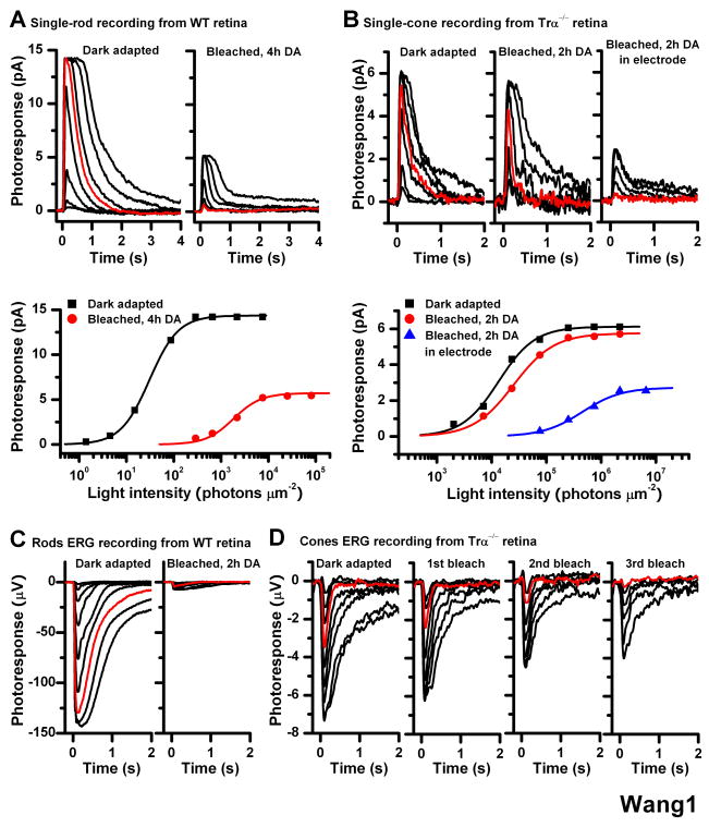
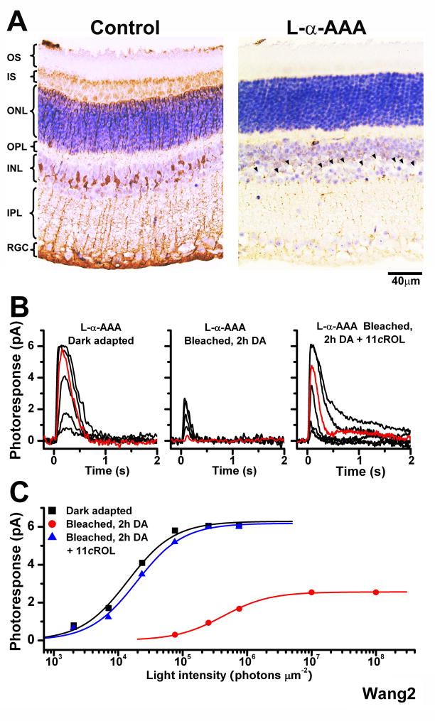
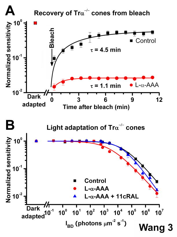
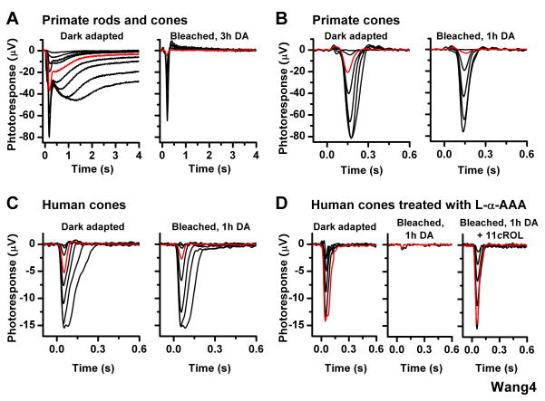
One of the fundamental mysteries of the human visual system is the continuous function of cone photoreceptors in bright daylight. As visual pigment is destroyed, or bleached, by light, cones require its rapid regeneration, which in turn involves rapid recycling of the pigment's chromophore. The canonical visual cycle for rod and cone pigments involves recycling of their chromophore from all-trans retinol to 11-cis retinal in the pigment epithelium, adjacent to photoreceptors. However, shortcomings of this pathway indicate the function of a second, cone-specific, mechanism for chromophore recycling. Indeed, biochemical and physiological studies on lower species have described a cone-specific visual cycle in addition to the long-known pigment epithelium pathway. Two important questions remain, however: what is the role of this pathway in the function of mammalian cones, and is it present in higher mammals, including humans? Here, we show that mouse, primate, and human neural retinas promote pigment regeneration and dark adaptation selectively in cones, but not in rods. This pathway supports rapid dark adaptation of mammalian cones and extends their dynamic range in background light independently of the pigment epithelium. This pigment-regeneration mechanism is essential for our daytime vision and appears to be evolutionarily conserved.


Figures

Figure 1
Figure 1
Effect of bleach on rod and cone single-cell responses in isolated mouse retina Single-cell suction recordings of flash intensity-response families from rods of wild type mice (A) and cones of Trα−/− mice (B). Top panels show test flash responses from individual cells recorded in dark-adapted state (left) or following a 40-s 500 nm light bleach and 4-h (rod) or 2-h (cone) dark recovery period (right). Right-most panel in (B) shows responses from a cone that was bleached and held in the recording electrode during the dark recovery period. Here and in all subsequent figures, photoresponses were generated by 20-ms test flashes delivered at time 0 and of intensity increasing in 0.5 log unit steps. Red traces represent photoresponses to 291 photons μm−2, 500 nm for rods (A) and to 75,814 photons μm−2, 500 nm for cones (B). Bottom panels show the corresponding intensity-response relations for these cells, fit with Michaelis-Menten function R/Rmax = I/(I+IO), where R/Rmax is the normalized response amplitude, I is the flash intensity, and IO is the intensity required to produce half-saturating response. The desensitization produced by the bleach was persistent in rods but largely reversed in cones from isolated retina in the absence of pigment epithelium (C) Rod ERG responses from isolated wild type mouse retina in darkness (left) and following a bleach and 2-h dark recovery period (right). Red traces represent photoresponses to 1,977 photons μm−2 500 nm (D) Cone ERG responses from Trα−/− retina in dark-adapted state (left) and after each of three subsequent bleaches, followed by dark recovery period (right three panels). The substantial desensitization induced by the bleach was persistent in rods but was largely reversed in cones Methods: Mice were dark adapted overnight, euthanized by CO2 asphyxiation, and the eyes enucleated under dim red light. Under infrared light, the eyeballs were hemisected, and the retinas were removed and placed in L-15 medium saturated with pure oxygen. To block Müller cells function, prior to isolating the retina the eyecup was incubated with 10 mM L-α-AAA dissolved in L-15 medium, pH 7.4, for 2.5 h in oxygen-saturated chamber at room temperature. Single-cell suction recordings for rods and cones were done as described previously [9, 19]. Rod and cone electroretinogram (ERG) photoresponses from isolated mouse retina were done as described previously [8, 20, 21]. By pharmacologically blocking synaptic transmission (see Supplemental Experimental Procedures), we recorded the photoreceptor component (a-wave) of isolated retina ERG responses. Test flashes at 500 nm were delivered from an optical bench using a set of calibrated neutral density filters. The signals were amplified, low-pass filtered at 30 Hz (8-pole Bessel) and digitized at 100 Hz for further analysis. Flash sensitivity was calculated from the linear region of the intensity-response curve as the ratio of response amplitude and flash intensity.
Figure 2
Figure 2
Effect of the Müller cell inhibitor L-α-AAA on the recovery of mouse cone sensitivity following a bleach (A) Comparison of the morphology of control retina (left) and retina incubated in 10 mM L-α-AAA gliotoxin for 2.5 h (right). Missing Müller cell nuclei in right panel are indicated by arrowheads (B) Cone suction recordings from isolated Trα−/− retina pretreated with 10 mM L-α-AAA in darkness for 2.5 h and then transferred to control solution prior to recordings. Cone test-flash responses from retina in dark-adapted state (left), bleached and incubated in darkness for 2 h (middle), and bleached and incubated in darkness for 2 h in the presence of 11-cis retinol (11cROL, right). Red traces represent photoresponses to 75,814 photons μm−2, 500 nm (C) Intensity-response relations of the cells from (B) fitted with Michaelis-Menten function. The recovery of sensitivity and amplitude of cones from isolated retina were blocked by the gliotoxin, but brought back by the addition of 11-cis retinol.
Figure 3
Figure 3
Effect of the retina visual cycle on mouse cone dark- and light adaptation (A) Recovery of cone sensitivity following a bleach at time 0 from ERG recordings of isolated Trα−/− retina in control solution (black, n = 5) and Trα−/− retina pre-treated for 2.5 h with 10 mM L-α-AAA (red, n = 5). The isolated Trα−/− retina promoted rapid cone dark adaptation at body temperature that could be inhibited by the gliotoxin L-α-AAA. Data are fitted by single exponential functions (B) Cone desensitization in background light from ERG recordings of isolated Tr-α−/− retina. Data are fitted with the Weber-Fechner relation S/SDA = (1 + IB/IO) 1, where S is the light-adapted sensitivity, SDA is the dark-adapted sensitivity, IB is the intensity of the background, and IO is the background that reduced sensitivity to half of SDA. IO was 25,165 photons μm−2 s−1 for cones in control retinas (n = 7) and was reduced to 2,747 photons μm−2 s−1 in L-α-AAA treated retinas (n = 8). Exogenous 11-cis retinal reversed the effect of gliotoxin, with IO = 20,749 photons μm −2 s−1 (n = 7). All sensitivity measurements were normalized to the corresponding dark-adapted value. Error bars give SEM.
Figure 4
Figure 4
Effect of bleach on cone ERG responses in isolated primate and human retinas (A) Primate rod (slow, high sensitivity) and cone (fast, low sensitivity) ERG responses from retina dark adapted in eyecup after enucleation in bright light (left). Following a subsequent bleach of the isolated retina, the rod component was abolished whereas the cone component recovered substantially (right) (B) Cone ERG responses from primate retina, dark adapted in the absence of pigment epithelium after enucleation in bright light (left) and following subsequent bleach and 1-h dark incubation (right). Primate cone response amplitude and sensitivity recovered substantially following the bleach (C) Cone ERG responses from human retina, dark adapted in the absence of pigment epithelium after enucleation in bright light (left) and following subsequent bleach and 1-h dark incubation (right). Human cone response amplitude and sensitivity recovered substantially following the bleach (D) Cone ERG recordings from human retina pretreated with 10 mM L-α-AAA in darkness for 2.5 h and then transferred to control solution prior to recordings. Cone test-flash responses from retina in dark-adapted state (left) and bleached and dark-adapted for 1 h in control solution (middle) or in the presence of 11-cis retinol (11cROL, right). The gliotoxin blocked recovery of sensitivity and amplitude of human cones from isolated retina, but exogenous 11-cis retinol reversed that effect. Red traces represent photoresponses to 18,560 photons μm−2 560 nm (A–C) and to 663,500 photons μm−2, 560 nm (D).

References

    1. Ebrey T, Koutalos Y. Vertebrate photoreceptors. Prog Retin Eye Res. 2001;20:49–94. - PubMed
    1. Saari JC. Biochemistry of visual pigment regeneration: the Friedenwald lecture. Invest Ophthalmol Vis Sci. 2000;41:337–348. - PubMed
    1. Mata NL, Radu RA, Clemmons RC, Travis GH. Isomerization and oxidation of vitamin A in cone-dominant retinas: a novel pathway for visual-pigment regeneration in daylight. Neuron. 2002;36:69–80. - PMC - PubMed
    1. Bustamante JJ, Ziari S, Ramirez RD, Tsin AT. Retinyl ester hydrolase and the visual cycle in the chicken eye. Am J Physiol. 1995;269:R1346–1350. - PubMed
    1. Das SR, Bhardwaj N, Kjeldbye H, Gouras P. Muller cells of chicken retina synthesize 11-cis-retinol. Biochem J. 1992;285(Pt 3):907–913. - PMC - PubMed

Publication types