. 2009 Sep 24;35(6):769-81.
doi: 10.1016/j.molcel.2009.07.030.

Reconstitution of heterochromatin-dependent transcriptional gene silencing

Affiliations

Reconstitution of heterochromatin-dependent transcriptional gene silencing

Aaron Johnson et al. Mol Cell. .

Abstract

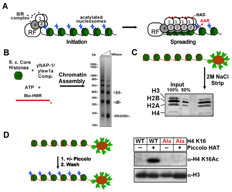
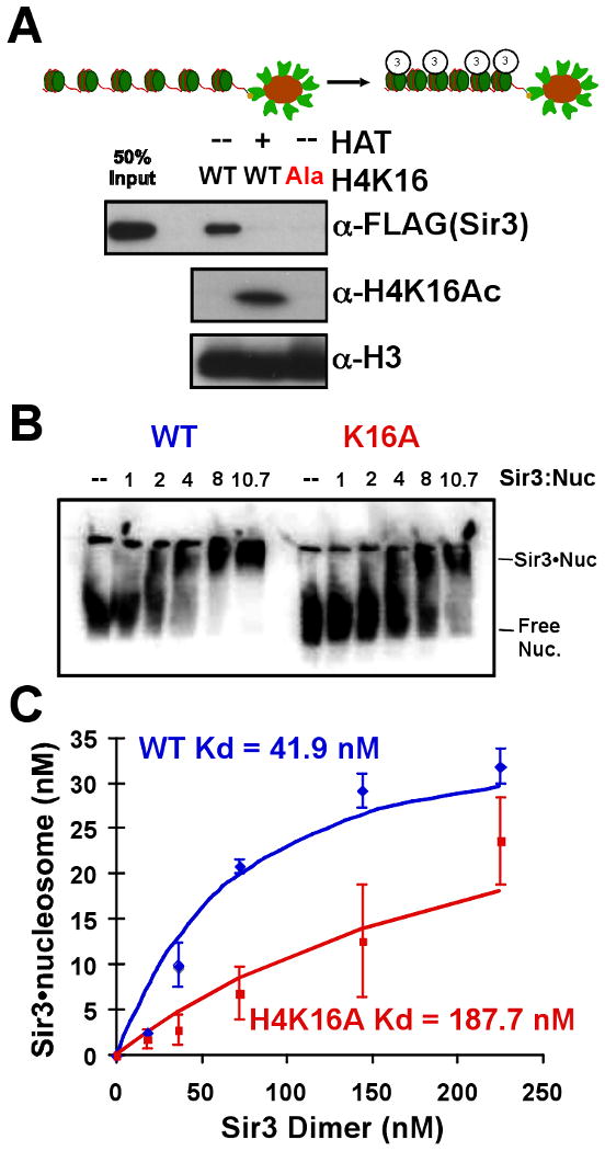
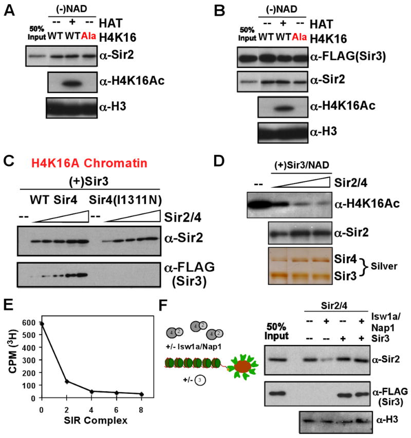
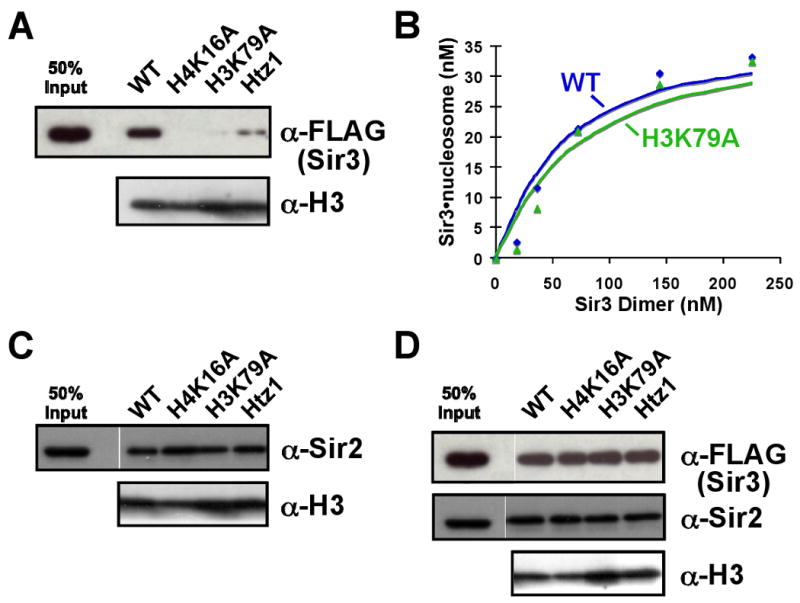
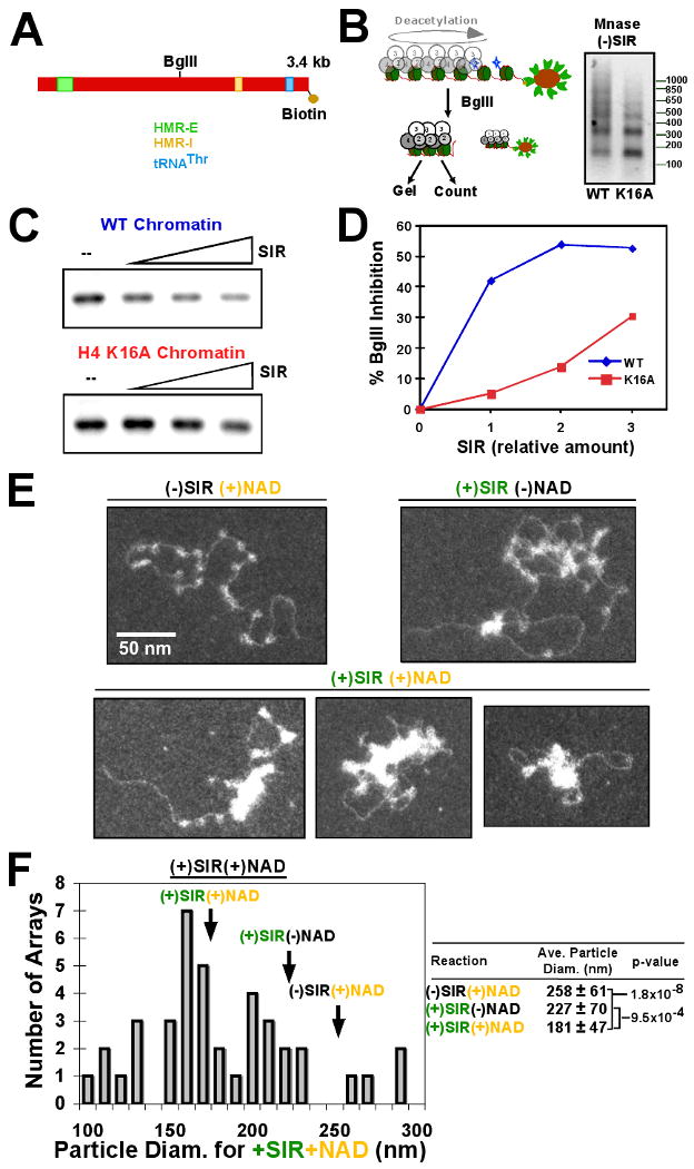
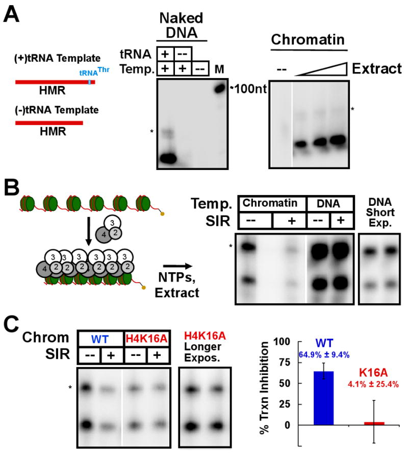
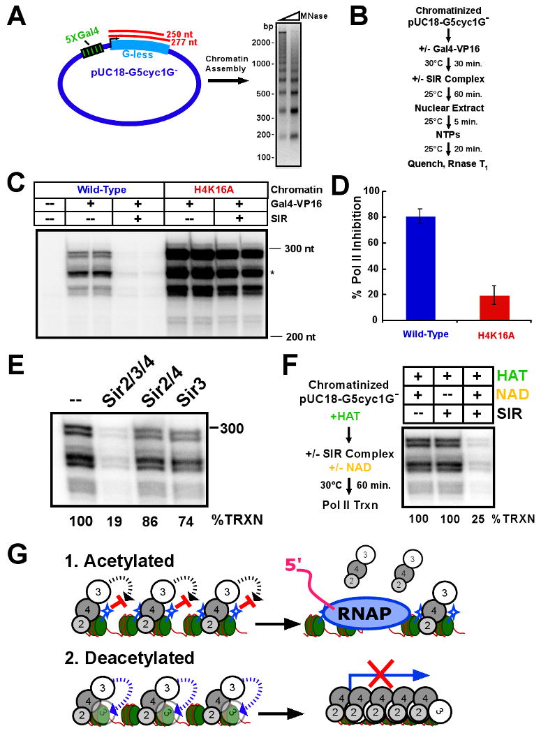
Heterochromatin assembly in budding yeast requires the SIR complex, which contains the NAD-dependent deacetylase Sir2 and the Sir3 and Sir4 proteins. Sir3 binds to nucleosomes containing deacetylated histone H4 lysine 16 (H4K16) and, with Sir4, promotes spreading of Sir2 and deacetylation along the chromatin fiber. Combined action of histone modifying and binding activities is a conserved hallmark of heterochromatin, but the relative contribution of each activity to silencing has remained unclear. Here, we reconstitute SIR-chromatin complexes using purified components and show that the SIR complex efficiently deacetylates chromatin templates and promotes the assembly of altered structures that silence Gal4-VP16-activated transcription. Silencing requires all three Sir proteins, even with fully deacetylated chromatin, and involves the specific association of Sir3 with deacetylated H4K16. These results define a minimal set of components that mediate heterochromatic gene silencing and demonstrate distinct contributions for histone deacetylation and nucleosome binding in the silencing mechanism.


Figures

Figure 1
Figure 1. Reconstitution of acetylated yeast chromatin
(A) A basic model for SIR complex assembly into silent chromatin. DNA-binding proteins act as recruitment factors (RF) to direct the SIR complex to target chromosomal regions. An iterative cycle of NAD-dependent histone deacetylation and direct chromatin association by the SIR complex leads to spreading of the SIR complex and produces a SIR-coated chromatin fiber that silences transcription. Deacetylation produces the small-molecule O-acetyl-ADP ribose (OAADPR or AAR), which may be incorporated into the SIR complex during assembly on chromatin. (B) Scheme for chromatin assembly using E. coli-expressed yeast histones, purified assembly factors, and a linear biotinylated HMR DNA fragment. Limited micrococcal nuclease (MNase) digestion of the assembled chromatin yields a ladder pattern indicating regularly-spaced nucleosomes. Positions of mono-, di-, and trinucleosomal DNA are indicated. (C) The biotinylated chromatin was immobilized on streptavidin-coated magnetic beads and the assembly factors were washed away. Histones remained stably associated in the proper stoichiometry after conjugation and washing. Input histones were stoichiometric by Coomassie, but each histone stains to a different extent by silver, shown in the panel. (D) The bead-conjugated chromatin is acetylated with the Piccolo HAT complex, which is subsequently washed away. Piccolo treatment produces a strong signal for acetylation of H4K16, which is abolished upon H4K16A mutation.
Figure 2
Figure 2. Sir3 binding to chromatin requires unmodified H4K16
Sir3 was incubated with bead-conjugated chromatin, the beads were washed, and chromatin association was determined by Western blot analysis of the bead-bound fraction. (A) Sir3 alone was incubated with wild-type chromatin (WT) +/- histone acetyltransferase treatment (HAT) or chromatin containing a H4K16 to alanine mutant (H4K16:Ala). (B) Sir3 association with a 32P-labeled mononucleosome was tested by gel shift assay. Nucleosomes with either wild-type (left) or H4K16A (right) histones were pre-incubated with Sir3 (ratio of Sir3 monomer: nucleosome shown) and subsequently run on a native polyacrylamide gel. The gel was dried and visualized by storage phosphor screen. (C) Quantification of bound vs. free nucleosome was used to determine an apparent Kd value for a mononucleosome and a dimer of Sir3.
Figure 3
Figure 3. Sir2/4 activities on chromatin
Sir2/4 (A) and the SIR complex (B) were used in chromatin association assays similar to those in Figure 2A, in the absence of NAD. (C) Titration of either wild-type or Sir4I1311N Sir2/4 subcomplex into a chromatin pull-down reaction containing a constant amount of Sir3 and H4K16A chromatin. (D) Sir2/4 was titrated into a reaction containing HAT-treated chromatin, Sir3, and NAD. The presence of Sir3 and Sir4 was determined by silver stain. (E) Bead-bound chromatin was acetylated in the presence of 3H-acetyl-CoA and subsequently treated with the SIR complex in the presence of NAD. Beads were washed and counted for 3H by liquid scintillation. (F) Sir2/4 binding to chromatin was assayed in the presence of the chromatin assembly components Isw1a and Nap1. The effect of addition of Sir3 was further tested.
Figure 4
Figure 4. Anti-silencing chromatin is refractory to Sir3 binding
Magnetic bead pull-down experiments were performed similarly to those in Figure 2. Sir3 alone (A), Sir2/4 (C) or the SIR complex (D) were incubated with wild-type chromatin, or chromatin containing either H4K16A, H3K79A, or the histone variant Htz1 replacing histone H2A. (B) Quantification of a mononucleosome gel-shift assay with either a wild-type or H3K79A nucleosome and Sir3.
Figure 5
Figure 5. NAD-dependent deacetylation by the SIR complex alters the structure of chromatin
(A) Diagram of the HMR-containing DNA template and scheme (B) for this assay. (right) The fragment released from the beads by BglII cleavage was digested by micrococcal nuclease. (C) Acetylated wild-type or H4K16A chromatin was analyzed as in the scheme. Reactions were quenched with EDTA and either run on an agarose gel and imaged by storage phosphor screen (C) or analyzed by liquid scintillation counting (D). (E) Desthiobiotinylated-chromatin was acetylated on magnetic beads and eluted with free biotin. Acetylated chromatin was incubated with NAD only, SIR complex without NAD, or SIR complex with NAD and subsequently imaged by electron microscopy. (F) Histogram of particle diameters from (+)SIR(+)NAD sample. Arrows indicate average particle diameter of indicated samples. (right) Table containing average particle diameters from EM experiments with standard deviations and p-values from two-tailed T-test analysis as indicated.
Figure 6
Figure 6. The SIR complex inhibits RNA Polymerase III transcription on chromatin
Transcription assays were performed using a whole-cell yeast extract enriched for transcription activity and RNA products were followed by α-32P-labeled UTP incorporation. Products were purified and separated on a 5 % acrylamide-urea-TBE gel and visualized on a storage phosphor screen. (A) The naked DNA template used in this study, a fragment of the HMR region including the flanking tRNA gene, was used in the transcription assay (left). Control reactions without template or with the DNA template truncated to remove the tRNA gene were also performed. (right) The chromatinized DNA was used as a template for extract-dependent transcription. (B) The SIR complex was pre-incubated with the chromatin template prior to initiation of transcription (scheme at left). Transcriptional repression of the SIR complex on the chromatin template was compared to an equal amount of the naked DNA template. The asterisk (*) indicates the unprocessed tRNA transcript. (C) Wild-type and H4K16A chromatin were compared in the ability to allow SIR-dependent repression of transcription. (right) Quantification by storage phosphor screen of three such experiments with standard deviations.
Figure 7
Figure 7. The SIR complex represses activator-dependent RNA Polymerase II transcription
(A) Diagram of a plasmid template bearing an array of Gal4 binding sites upstream of a CYC1 promoter-driven G-less cassette with two expected transcripts, 250 and 277 nt in length (left). The plasmid was assembled into chromatin and analyzed as in Figure 1B for chromatinization of the DNA by limited micrococcal nuclease digestion. (B) Scheme of the activator-dependent transcription experiment. (C) Chromatinized G-less cassette plasmid, assembled with either wild-type or H4K16A histone octamer, was incubated with or without the SIR complex before initiation of transcription. RNase T1-treated products from duplicate reactions were resolved by denaturing PAGE. Migration positions of 300 and 200 nt RNA markers is shown. The asterisk marks the larger of the two expected transcripts. The topmost doublet band was most likely a read-through transcription product from the G-less region. (D) Quantification of transcription inhibition by the SIR complex on either wild-type or H4K16A templates by storage phosphor screen analysis. Shown are the averages of three experiments with standard deviations. (E) Individual components of the SIR complex were tested for Pol II repression as in (C). (F) Chromatinized G-less cassette plasmid was acetylated prior to SIR complex incubation performed in the absence or presence of NAD (scheme at left). Transcription assays were performed and products analyzed as above (right). (G) Model for transcriptional silencing by Sir3 lockdown onto chromatin, mediated by Sir4 and deacetylated H4K16. 1. Acetylated chromatin allows Sir2/4 binding, but prevents direct association of Sir3 with nucleosomes. This loosely associated SIR complex is permissive to transcription. 2. NAD-dependent deacetylation generates a high-affinity substrate for Sir3 lockdown onto chromatin and promotes an altered SIR-chromatin complex that is repressive to transcription.

References

    1. Altaf M, Utley RT, Lacoste N, Tan S, Briggs SD, Cote J. Interplay of chromatin modifiers on a short basic patch of histone H4 tail defines the boundary of telomeric heterochromatin. Mol Cell. 2007;28:1002–1014. - PMC - PubMed
    1. Buchberger JR, Onishi M, Li G, Seebacher J, Rudner AD, Gygi SP, Moazed D. Sir3-nucleosome interactions in spreading of silent chromatin in Saccharomyces cerevisiae. Mol Cell Biol. 2008;28:6903–6918. - PMC - PubMed
    1. Chang JF, Hall BE, Tanny JC, Moazed D, Filman D, Ellenberger T. Structure of the coiled-coil dimerization motif of Sir4 and its interaction with Sir3. Structure. 2003;11:637–649. - PubMed
    1. Chou CC, Li YC, Gartenberg MR. Bypassing Sir2 and O-acetyl-ADP-ribose in transcriptional silencing. Mol Cell. 2008;31:650–659. - PMC - PubMed
    1. Dorigo B, Schalch T, Bystricky K, Richmond TJ. Chromatin fiber folding: requirement for the histone H4 N-terminal tail. J Mol Biol. 2003;327:85–96. - PubMed

Publication types

MeSH terms

Substances

LinkOut - more resources