. 2010 Jan;138(1):347-59.
doi: 10.1053/j.gastro.2009.09.015. Epub 2009 Sep 24.

Modulation of hepatic fibrosis by c-Jun-N-terminal kinase inhibition

Affiliations

Modulation of hepatic fibrosis by c-Jun-N-terminal kinase inhibition

Johannes Kluwe et al. Gastroenterology. 2010 Jan.

Abstract

Background & aims: c-Jun N-terminal kinase (JNK) is activated by multiple profibrogenic mediators; JNK activation occurs during toxic, metabolic, and autoimmune liver injury. However, its role in hepatic fibrogenesis is unknown.

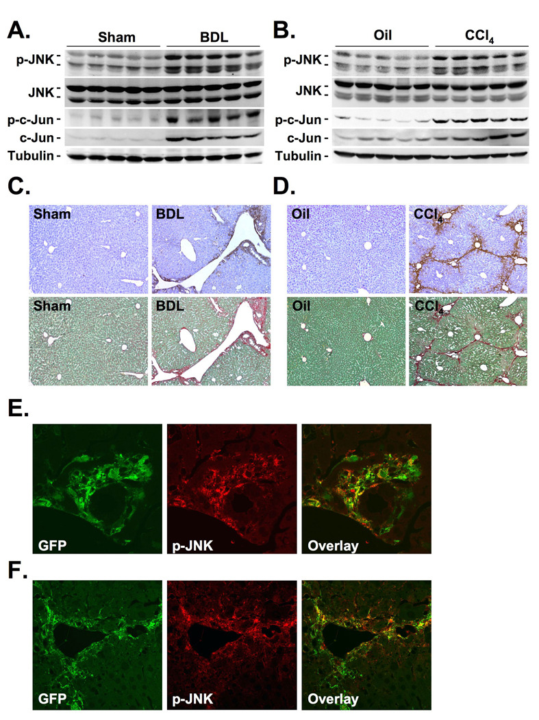
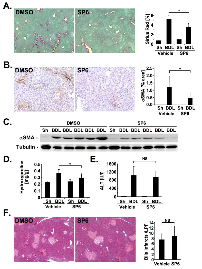
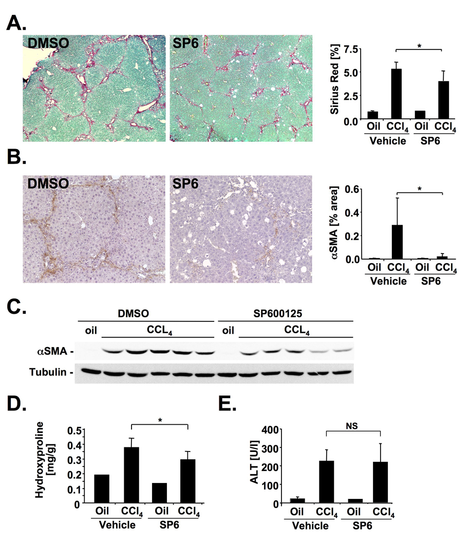
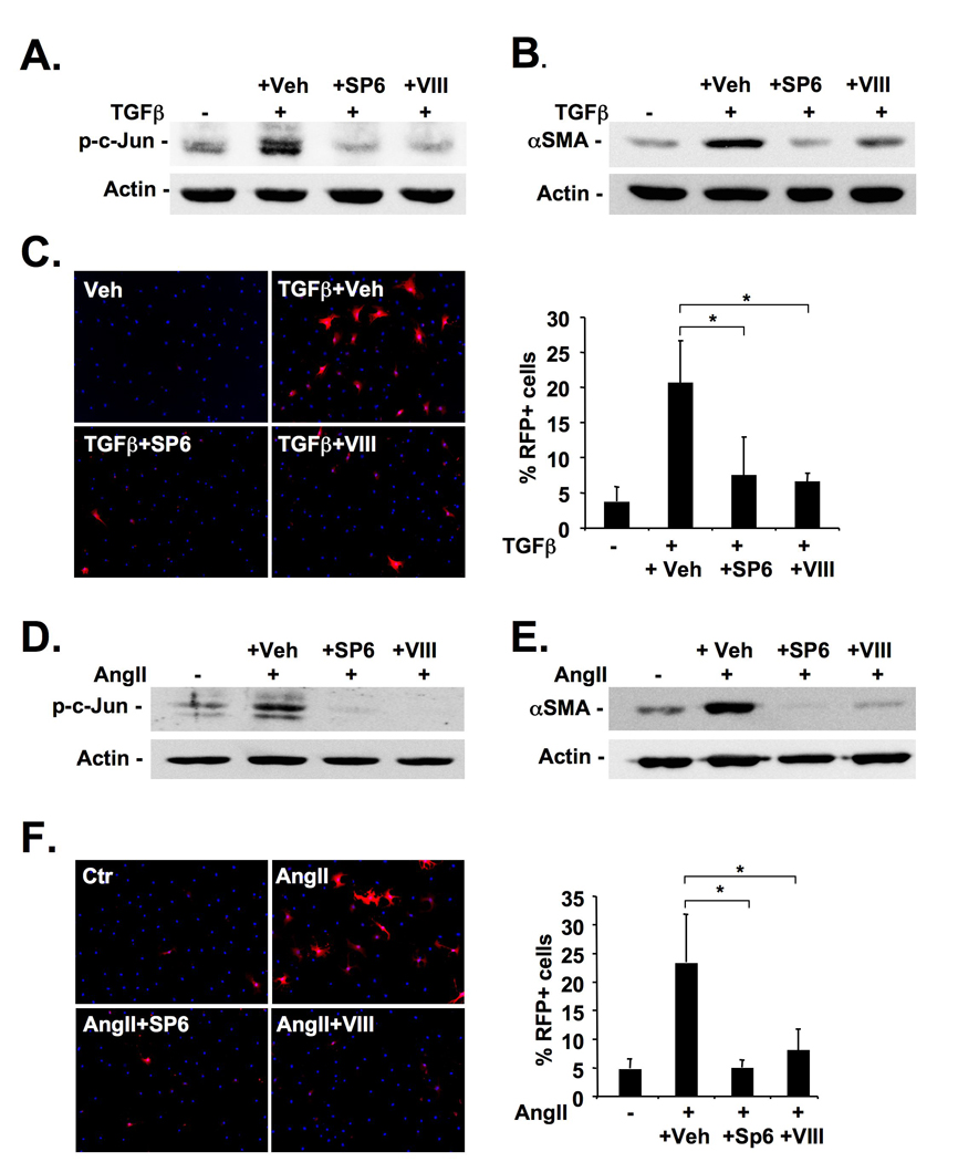
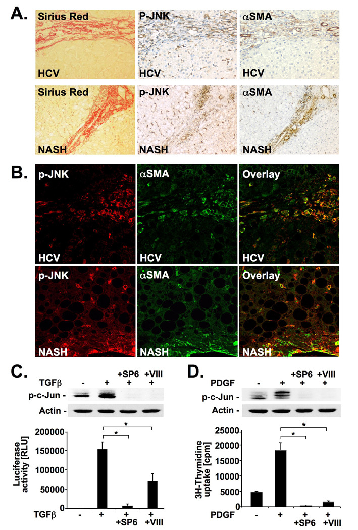
Methods: JNK phosphorylation was detected by immunoblot analysis and confocal immunofluorescent microscopy in fibrotic livers from mice after bile duct ligation (BDL) or CCl(4) administration and in liver samples from patients with chronic hepatitis C and non-alcoholic steatohepatitis. Fibrogenesis was investigated in mice given the JNK inhibitor SP600125 and in JNK1- and JNK2-deficient mice following BDL or CCl(4) administration. Hepatic stellate cell (HSC) activation was determined in primary mouse HSCs incubated with pan-JNK inhibitors SP600125 and VIII.

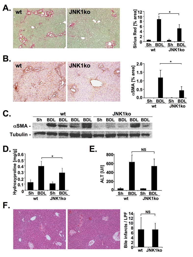
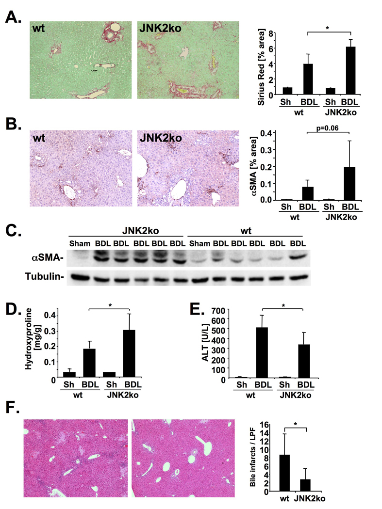
Results: JNK phosphorylation was strongly increased in livers of mice following BDL or CCl(4) administration as well as in human fibrotic livers, occurring predominantly in myofibroblasts. In vitro, pan-JNK inhibitors prevented transforming growth factor (TGF) beta-, platelet-derived growth factor-, and angiotensin II-induced murine HSC activation and decreased platelet-derived growth factor and TGF-beta signaling in human HSCs. In vivo, pan-JNK inhibition did not affect liver injury but significantly reduced fibrosis after BDL or CCl(4). JNK1-deficient mice had decreased fibrosis after BDL or CCl(4), whereas JNK2-deficient mice displayed increased fibrosis after BDL but fibrosis was not changed after CCl(4). Moreover, patients with chronic hepatitis C who displayed decreased fibrosis in response to the angiotensin receptor type 1 blocker losartan showed decreased JNK phosphorylation.

Conclusions: JNK is involved in HSC activation and fibrogenesis and represents a potential target for antifibrotic treatment approaches.


Conflict of interest statement

Disclosures: No conflicting interests exist.

Figures

Figure 1
Figure 1. JNK activation during hepatic fibrogenesis occurs in myofibroblasts
A–D. Balb/c mice underwent BDL or sham operation for 15 days, or 8 gavages of oil or CCl4 (0.5 µl/g). JNK activation was determined by immunoblotting of phospho-c-Jun or p-JNK (A–B.). Serial sections were stained for p-JNK and by picrosirius red (C–D.). E–F. Collagen-GFP mice underwent BDL or sham operation for 15 days (E.), or were treated with 8 injections of CCl4 or oil (F.). Sections were stained for GFP (green staining) and p-JNK (red staining) followed by confocal microscopy.
Figure 2
Figure 2. JNK inhibition ameliorates bile duct ligation-induced hepatic fibrosis
A–D. Male Balb/c mice underwent BDL for 15 days, and were treated with 1 mg SP600125 twice daily (n=7) or DMSO vehicle (n=10). Hepatic fibrosis was evaluated by Sirius Red staining (A), αSMA immunohistochemistry (B), αSMA immunoblotting (C), and hydroxyproline content (D). E.–F. Hepatic injury in SP600125- (n=7) or vehicle-treated (n=8) mice was determined by ALT levels (E) and bile infarcts 5 days after BDL (F). * p<0.05
Figure 3
Figure 3. JNK inhibition ameliorates CCl4-induced hepatic fibrosis
A–E. SP600125- treated (n=9) and vehicle-treated (n=9) Balb/c mice received 4 gavages of CCl4. Hepatic fibrosis and injury were evaluated by Sirius Red staining (A), αSMA immunohistochemistry (B), αSMA immunoblotting (C), hydroxyproline content (D) and ALT levels 48h after the last CCl4 injection (E). * p<0.05
Figure 4
Figure 4. JNK promotes hepatic stellate activation and proliferation
A–B. Primary mouse HSCs were pretreated with SP600125 (5 µM), inhibitor VIII (16 µM) or vehicle (0.1% DMSO) followed by TGFβ (2 ng/ml) treatment. c-Jun phosphorylation (A) and αSMA expression (B) were determined by immunoblot 60 minutes and 48h after TGFβ treatment, respectively. C. HSCs were isolated from αSMA-RFP mice, pretreated with JNK inhibitors or vehicle for 24h and treated with TGFβ (2 ng/ml) for 48h followed by Hoechst 33342 staining and quantification of RFP expression. D–E. One day after isolation, primary mouse HSCs were pretreated with JNK inhibitors or vehicle (see above) followed by treatment with AngII (10−7 M). c-Jun phosphorylation (D) and αSMA expression (E) were determined by immunoblot 20 minutes and 48h after AngII treatment, respectively. F. HSCs from αSMA-RFP mice were pretreated as described above followed by treatment with AngII for 48h and evaluation of RFP expression.
Figure 5
Figure 5. JNK1 promotes hepatic fibrosis
A.–D. JNK1-deficient (n=10) or control mice (n=7) underwent BDL for 21 days. Hepatic fibrosis was evaluated by Sirius Red staining (A), αSMA immunohistochemistry (B), αSMA immunoblotting (C), and hydroxyproline content (D). E–F. Hepatic injury was quantified by ALT levels (E) and bile infarcts (f) in JNK1-deficient (n=9) and control mice (n=9) five days after BDL. * p<0.05
Figure 6
Figure 6. JNK2 decreases hepatic fibrosis
A–D. JNK2-deficient mice (n=9) or C57Bl/6 control mice (n=10) underwent BDL for 21 days. Hepatic fibrosis was evaluated by Sirius Red staining (A), αSMA immunohistochemistry (B), αSMA immunoblotting (C), and hydroxyproline content (D). E–F. Hepatic injury was quantified by ALT levels (E.) and bile infarcts (F.) in JNK2-deficient (n=8) and C57Bl/6 control mice (n=6) five days after BDL. * p<0.05
Figure 7
Figure 7. JNK is activated in human liver fibrosis and promotes TGFβ and PDGF signaling in human hepatic stellate cells
A–B. Serial sections of fibrotic livers from patients with HCV (n=3) or NASH (n=3) were stained for p-JNK, αSMA or by picrosirius red. Single sections were stained for αSMA (green) and p-JNK (red) followed by confocal microscopy. Shown are representative images from one patient each (B). C. Primary human HSCs were pretreated with SP600125 or JNK inhibitor VIII. c-Jun phosphorylation was determined by immunoblot 60 minutes after TGFβ (2 ng/ml) treatment (upper panel). (CAGA)9-driven luciferase was determined 6 hours after TGFβ (10 pg/ml) (lower panel). D. Primary human HSCs were pretreated with JNK inhibitors followed by PDGF (20 ng/ml) treatment. Phosphorylation of c-Jun (upper panel) and [3H]-thymidine incorporation (lower panel) were determined 15 minutes and 26h after PDGF stimulation, respectively. * p<0.05

Similar articles

Cited by

References

    1. Bataller R, Brenner DA. Liver fibrosis. J Clin Invest. 2005;115:209–218. - PMC - PubMed
    1. Friedman SL. Mechanisms of hepatic fibrogenesis. Gastroenterology. 2008;134:1655–1669. - PMC - PubMed
    1. Minden A, Karin M. Regulation and function of the JNK subgroup of MAP kinases. Biochim Biophys Acta. 1997;1333:F85–F104. - PubMed
    1. Weston CR, Davis RJ. The JNK signal transduction pathway. Curr Opin Cell Biol. 2007;19:142–149. - PubMed
    1. Hibi M, Lin A, Smeal T, Minden A, Karin M. Identification of an oncoprotein- and UV-responsive protein kinase that binds and potentiates the c-Jun activation domain. Genes Dev. 1993;7:2135–2148. - PubMed

Publication types

MeSH terms