. 2009 Dec;37(12):1400-10.
doi: 10.1016/j.exphem.2009.09.006. Epub 2009 Sep 24.

Bone marrow engraftment but limited expansion of hematopoietic cells from multipotent germline stem cells derived from neonatal mouse testis

Affiliations

Bone marrow engraftment but limited expansion of hematopoietic cells from multipotent germline stem cells derived from neonatal mouse testis

Momoko Yoshimoto et al. Exp Hematol. 2009 Dec.

Abstract

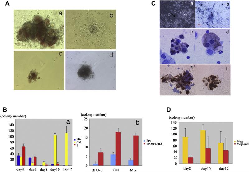
Objective: Multipotent germline stem (mGS) cells derived from neonatal mouse testis, similar to embryonic stem (ES) cells, differentiate into various types of somatic cells in vitro and produce teratomas after inoculation into mice. In the present work, we examined mGS cells for hematopoietic progenitor potential in vitro and in vivo.

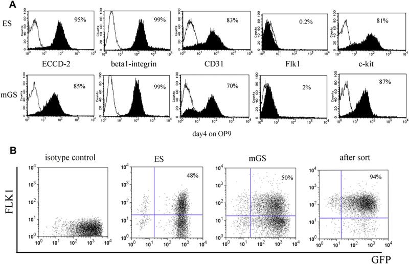
Materials and methods: mGS cells were differentiated on OP9 stromal cells and induced into Flk1(+) cells. Flk1(+) cells were sorted and replated on OP9 stromal cells with various cytokines and emerging hematopoietic cells were analyzed for lineage marker expression by fluorescein-activated cell sorting, progenitor activity by colony assay, and stem cell transplantation assay.

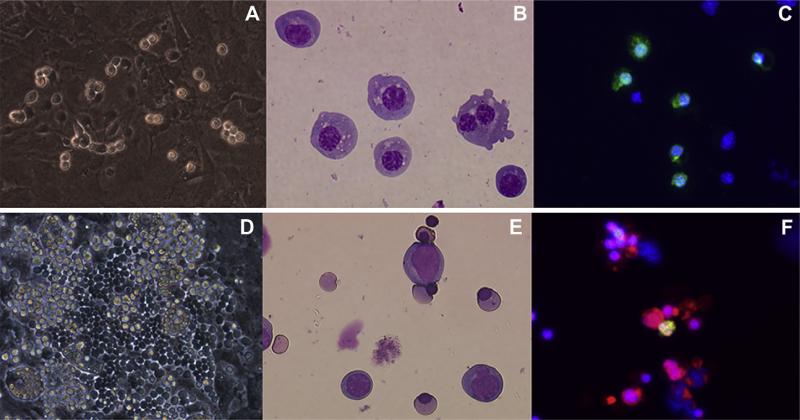
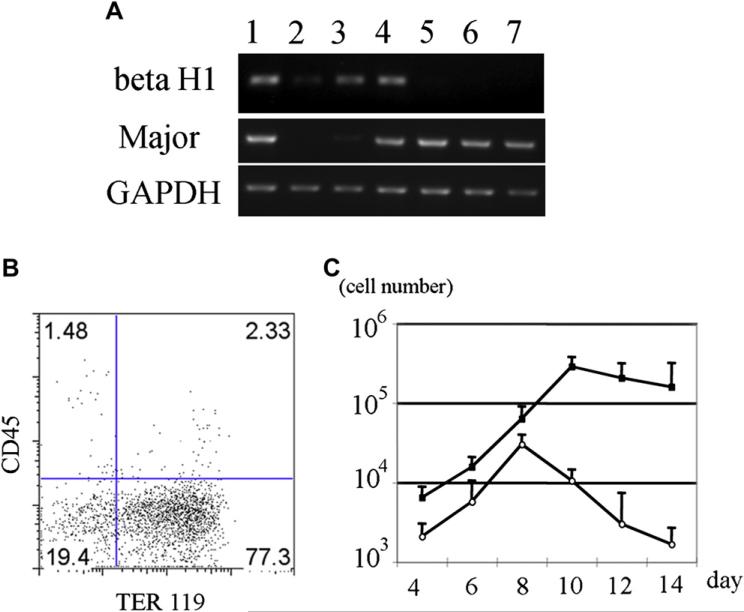
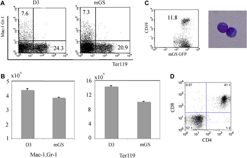
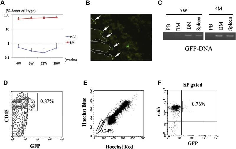
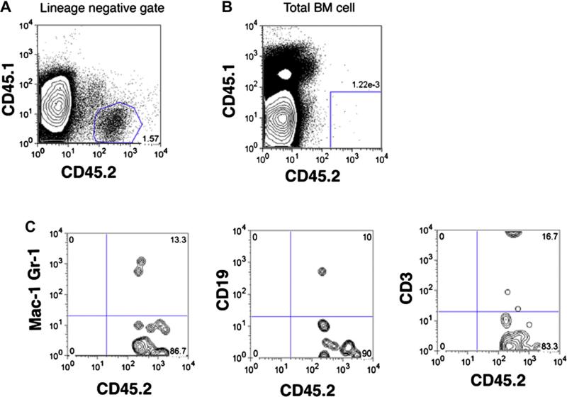
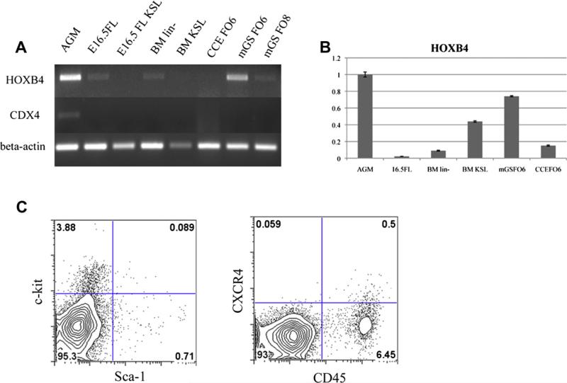
Results: mGS cells, like ES cells, produce hematopoietic progenitors, including both primitive and definitive erythromyeloid, megakaryocyte, and B- and T-cell lineages via Flk1(+) progenitors. When transplanted into the bone marrow (BM) of nonobese diabetic/severe combined immunodeficient (NOD/SCID) gammac(null) mice directly, mGS-derived green fluorescent protein (GFP)-positive cells were detected 4 months later in the BM and spleen. GFP(+) donor cells were also identified in the Hoechst33342 side population, a feature of hematopoietic stem cells. However, these mGS-derived hematopoietic cells did not proliferate in vivo, even after exposure to hematopoietic stressors, such as 5-fluorouracil (5FU) injection or serial transplantation.

Conclusion: mGS cells produced multipotent hematopoietic progenitor cells with myeloid and lymphoid lineage potential in vitro and localized in the BM after intra-BM injection but, like ES cells, failed to expand or show stem cell repopulating ability in vivo.


Figures

Figure 1
Figure 1
Surface markers of mGS and ES cells. Surface markers of undifferentiated mGS cells (A, lower panel) are similar to those of ES cells (A, upper panel). mGS cells were induced on OP9 stromal cells for 4 days and were confirmed to express Flk1 (B).
Figure 2
Figure 2
Primitive and definitive erythroid cells are differentiated from mGS–derived Flk1+ cells. mGS-derived Flk1+ cells were cultured on OP9 cells and small round cells and cobblestone-forming areas were found [(A) day 3, (D) day 9). May-Giemsa staining of a cytospin preparation [(B); day 3, (E); day 9)]. Immunostaining of E1 antigen and Ter119 (C,F). (C) E1 antigen; green. (F) E1 antigen; green Ter119; red. Nuclear staining with Hoechst 33324; blue. Magnification (A,D): ×100; (B,C,E,F): ×200.
Figure 3
Figure 3
Erythropoiesis from mGS Flk1+ cells shows two different waves. Flk1+ cells were cultured on OP9 with erythropoietin (EPO) and with or without ACK45. The nonadherent cells in the culture were collected every other day, counted the number (C), examined the surface markers (B) and hemoglobin gene expression (A). Sequential RT-PCR analysis shows expression of β H1 hemoglobin mRNA, followed by b-major hemoglobin mRNA expression (A). E12.5 fetal liver (lane 1), E8.0 embryo (lane 2), mGS- Flk1+ cells on OP9 for 4 days (lane 3), for 6 days (lane 4), for 8 days (lane 5), for 10 days (lane 6), for 12 days (lane 7). Flk1+ cells were cultured on OP9 cells with EPO and with (white circle) or without ACK45 (black square) (C). Most cells in the culture were Ter119+ erythrocyte (B). Definitive erythropoiesis was blocked by ACK45 (C). GAPDH = glyceraldehydes phosphate dehydrogenase.
Figure 4
Figure 4
Myelolymphoid potential of mGS cells. Mac1+Gr1+, Ter119+, CD19+, and CD4+CD8+ cells were differentiated from mGS-derived Flk-1+ cells within OP9 culture (A,B,C) or OP9-DL1 culture (D). For Mac1+Gr1+, Ter119+ cells, cells were collected from 8–10 days culture (A,B). The numbers of myeloid and erythroid cells differentiated from mGS and ES cells were similar (B). For CD19+ (C), and CD4+CD8+ cells (D), cells were collected from 14–21 days culture. These FACS data are representative among three independent experiments.
Figure 5
Figure 5
Colony-forming ability and megakaryocyte potential of mGS Flk1+-derived cells. mGS-derived Flk-1+ cells produce hematopoietic progenitor cells that can form various kinds of colonies [(Aa) mixed colony, (Ab) granulocyte-macrophage (GM) colony, (Ac) burst-forming unit erythroid (BFU-E), (Ad) mast cell colony]. Mixed colonies were predominant when mGS-derived Flk1+ cells were cultured on OP9 cells for 4 to 6 days (Ba). Blue bar: mixed colony, yellow bar: GM colony, red bar: Erythroid colony. The number of all kinds of colonies was increased when mGS Flk1+ cells were cultured on OP9 with thrombopoietin (TPO) and interleukin (IL)-6 (Bb). Blue bar: with erythropoietin (EPO), red bar: with TPO and IL6. Megakaryocyte or proplatelet mGS-derived Flk1+ cells were observed within OP9 culture for 8–12 days. Proplatelet like cells (arrow Ca), Immunostaining of cultured cells with CD41 antibody secondary detected by alkaline phosphatase–conjugated antibody (Cb blue). Immunostaining of a cytospin preparation with CD41 antibody secondary detected by peroxidase conjugated antibody (Cc, Cd brown). After transferring cultured cells into Megacult conditions, megakaryocyte colonies (Cf) include mega-mix colonies (Ce) were found. These cells expressed acethylcholinestrase as evidenced by brown staining. The CFU-Mega was maintained during culturing day 8 to 12 in the presence of TPO and stem cell factor (D). Yellow bar: Megakaryo-colony, red bar: Mega-Mix colony. Magnification: (Ca, Cb) ×100; (Cc, Cd) ×200; (Ce, Cf) ×100.
Figure 6
Figure 6
Transplanted hematopoietic cells from GFP+ mGS cells can be detected in bone marrow (BM) 4 months after transplantation and displays stem cell phenotype. After transplantation, peripheral blood (PB) was analyzed every 4 weeks (A). BM cells from recipient mice 4 months after transplantation were analyzed by LSR II (D, E, F). GFP+CD45+ cells were detected (D). When the BM cells of the recipient mice were stained with Hoechst 33324, GFP+ cells were detected in the SP region (E, F). In the section of recipient BM, GFP+ cells were found attached to the endosteal region (B, arrow). RT-PCR shows donor-derived DNA in the BM and spleen at 7 weeks and 4 months after transplantation (C).
Figure 7
Figure 7
Analysis of primary recipient mice transplanted with mGS–derived hematopoietic cells. There are small percentages of mGS-derived cells in lineage negative fraction in the recipient bone marrow (BM) cells (A). When analyzing total recipient BM cells, mGS-derived cell can be detectable in very small percentage (B), but showed multi-lineage cell types (C).
Figure 8
Figure 8
mGS–derived cells express HOXB4, but not CDX4 or CXCR4. RT-PCR for CDX4 and HOXB4 among various hematopoitic cells (A). KSL = c-kit+Sca-1+lineage cells, BM lin = bone marrow lineage-negative cells, FO6 = Flk1 culture on OP9 day 6, FO8 = Flk1 culture on OP9 day 8. Relative HOXB4 expression in the various hematopoitic cells (B). With AGM as a reference, the relative expressions of HOXB4 in each sample were follows; 16.5FL 0.02, BM lin 0.09, BM KSL 0.43, mGS FO6 0.74, CCE FO6 0.15. c-kit, Sca-1, and CXCR4 expression on mGS-derived cells [(C) 6 days culture].

References

    1. Kyba M, Perlingeiro RC, Daley GQ. HoxB4 confers definitive lymphoid-myeloid engraftment potential on embryonic stem cell and yolk sac hematopoietic progenitors. Cell. 2002;109:29–37. - PubMed
    1. Wang Y, Yates F, Naveiras O, Ernst P, Daley GQ. Embryonic stem cell-derived hematopoietic stem cells. Proc Natl Acad Sci U S A. 2005;102:19081–19086. - PMC - PubMed
    1. Kanatsu-Shinohara M, Inoue K, Lee J, et al. Generation of pluripotent stem cells from neonatal mouse testis. Cell. 2004;119:1001–1012. - PubMed
    1. Kennedy M, Firpo M, Choi K, et al. A common precursor for primitive erythropoiesis and definitive haematopoiesis. Nature. 1997;386:488–493. - PubMed
    1. Kataoka H, Takakura N, Nishikawa S, et al. Expressions of PDGF receptor alpha, c-Kit and Flk1 genes clustering in mouse chromosome 5 define distinct subsets of nascent mesodermal cells. Dev Growth Differ. 1997;39:729–740. - PubMed

Publication types

MeSH terms

Substances