Review
. 2009 Dec;32(12):638-47.
doi: 10.1016/j.tins.2009.08.002. Epub 2009 Sep 24.

Molecular dissection of reactive astrogliosis and glial scar formation

Affiliations
Review

Molecular dissection of reactive astrogliosis and glial scar formation

Michael V Sofroniew. Trends Neurosci. 2009 Dec.

Abstract

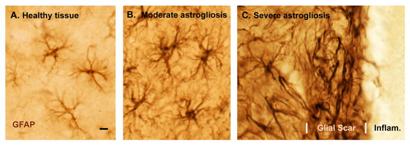
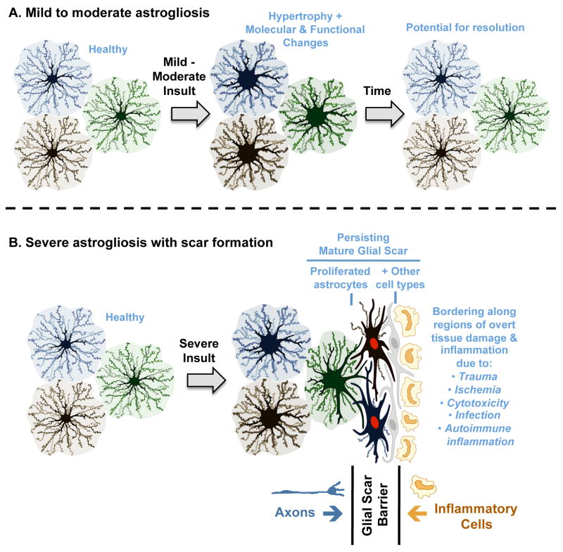
Reactive astrogliosis, whereby astrocytes undergo varying molecular and morphological changes, is a ubiquitous but poorly understood hallmark of all central nervous system pathologies. Genetic tools are now enabling the molecular dissection of the functions and mechanisms of reactive astrogliosis in vivo. Recent studies provide compelling evidence that reactive astrogliosis can exert both beneficial and detrimental effects in a context-dependent manner determined by specific molecular signaling cascades. Reactive astrocytes can have both loss of normal functions and gain of abnormal effects that could feature prominently in a variety of disease processes. This article reviews developments in the signaling mechanisms that regulate specific aspects of reactive astrogliosis and highlights the potential to identify novel therapeutic molecular targets for diverse neurological disorders.


Figures

Figure 1
Figure 1. Photomicrographs of astrocytes in healthy tissue and of different gradations of reactive astrogliosis and glial scar formation after tissue insults of different types and different severity
(A-C) Immunohistochemical staining of glial fibrillary protein (GFAP) in wild-type mice. Note that GFAP staining demonstrates the main stem processes and general appearance of the astrocytes, but does not reveal all of the fine branches and ramifications. (A) Appearance of ‘normal’ astrocytes in healthy cerebral cortex of an untreated mouse. Note that the territories of astrocyte processes do not overlap. (B) Moderately reactive astrogliosis in mouse cerebral cortex in response to intracerebral injection of the bacterial antigen, lipopolysaccharide (LPS). Note that the territories of moderately reactive astrocyte processes also do not overlap. (C) Severely reactive astrogliosis and glial scar formation adjacent to a region of severe traumatic injury and inflammation (Infam.) in the cerebral cortex. Note the extensive overlap and inter-digitations of processes of severely reactive and scar-forming astrocytes. Scale bar = 8μm.
Figure 2
Figure 2. Schematic representations of different gradations of reactive astrogliosis that vary with insult severity
(A) Mild to moderate reactive astrogliosis comprises variable changes in molecular expression and functional activity together with variable degrees of cellular hypertrophy. Such changes occur after mild trauma or at sites distant from a more severe injury, or after moderate metabolic or molecular insults or milder infections or inflammatory activation. These changes vary with insult severity, involve little anatomical overlap of the processes of neighboring astrocytes and exhibit the potential for structural resolution if the triggering insult is removed or resolves. (B) Severe reactive astrogliosis with persisting scar formation generally occurs along borders to areas of overt cell and tissue damage and inflammation. Glial scar formation includes newly proliferated astrocytes (with red nuclei in figure) and other cell types (with grey nuclei in figure) such as fibromeningeal cells and other glia. In the mature glial scar, astrocytes no longer occupy discrete domains and instead have overlapping processes. Mature glial scars tend to persist for long periods and act as barriers not only to axon regeneration but also to inflammatory cells in a manner that protects healthy tissue from nearby areas of intense inflammation.

References

    1. Barres BA. The mystery and magic of glia: a perspective on their roles in health and disease. Neuron. 2008;60:430–440. - PubMed
    1. Nedergaard M, et al. New roles for astrocytes: redefining the functional architecture of the brain. Trends Neurosci. 2003;26:523–530. - PubMed
    1. Pellerin L, et al. Activity-dependent regulation of energy metabolism by astrocytes: an update. Glia. 2007;55:1251–1262. - PubMed
    1. Seifert G, et al. Astrocyte dysfunction in neurological disorders: a molecular perspective. Nat Rev Neurosci. 2006;7:194–206. - PubMed
    1. Eddleston M, Mucke L. Molecular profile of reactive astrocytes -implications for their role in neurological disease. Neuroscience. 1993;54:15–36. - PMC - PubMed

Publication types

Substances