

Thermosensitive injectable hyaluronic acid hydrogel for adipose tissue engineering
- PMID: 19783043
- PMCID: PMC2783716
- DOI: 10.1016/j.biomaterials.2009.08.058
Thermosensitive injectable hyaluronic acid hydrogel for adipose tissue engineering
Abstract
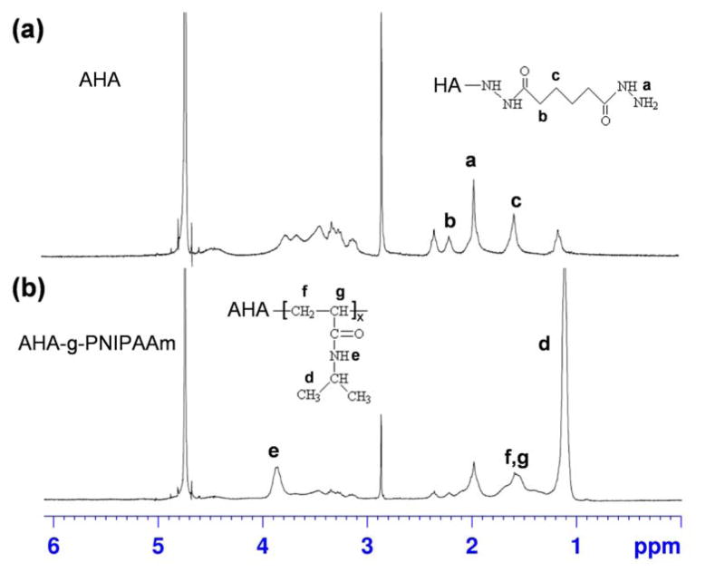
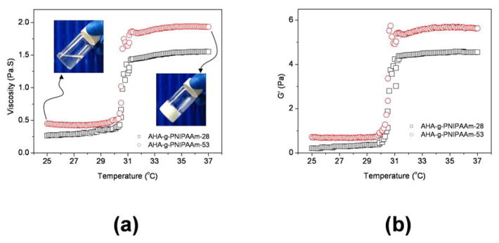
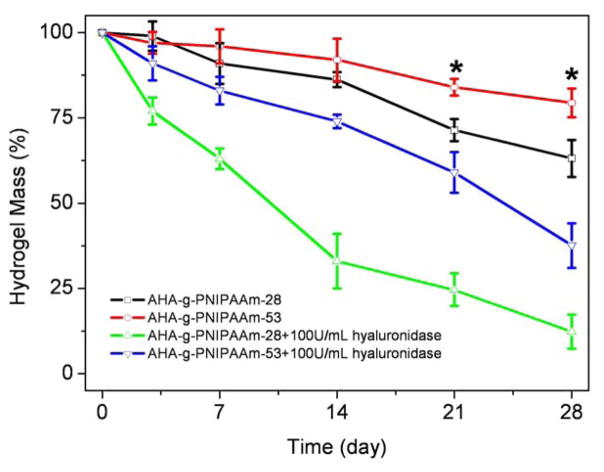
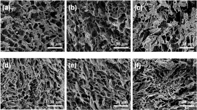
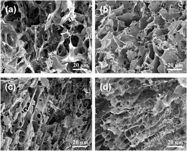
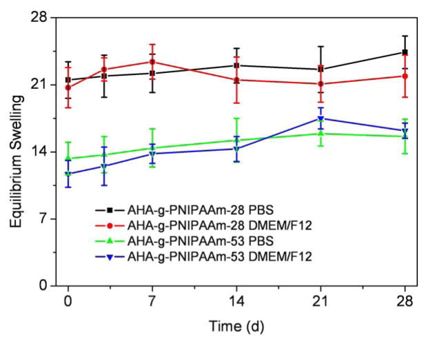
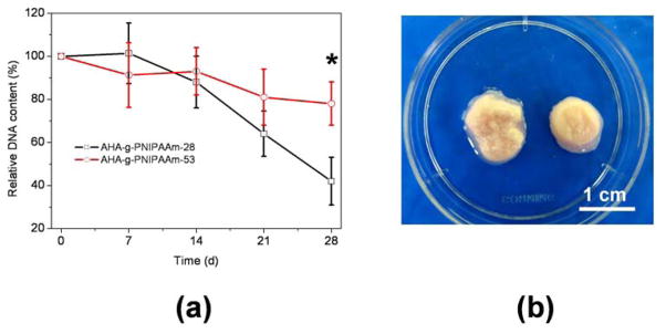
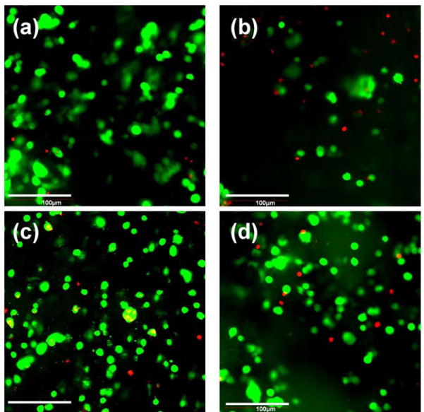
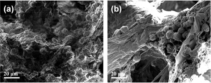
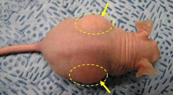
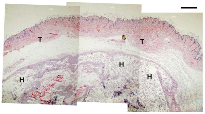
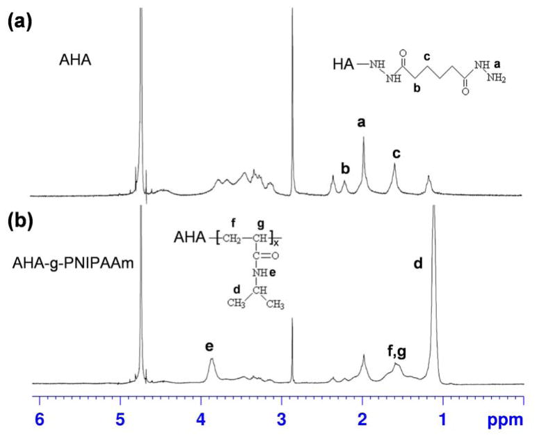
A series of thermosensitive copolymer hydrogels, aminated hyaluronic acid-g-poly(N-isopropylacrylamide) (AHA-g-PNIPAAm), were synthesized by coupling carboxylic end-capped PNIPAAm (PNIPAAm-COOH) to AHA through amide bond linkages. AHA was prepared by grafting adipic dihydrazide to the HA backbone and PNIPAAm-COOH copolymer was synthesized via a facile thermo-radical polymerization technique by polymerization of NIPAAm using 4,4'-azobis(4-cyanovaleric acid) as an initiator, respectively. The structure of AHA and AHA-g-PNIPAAm copolymer was determined by (1)H NMR. Two AHA-g-PNIPAAm copolymers with different weight ratios of PNIPAAm on the applicability of injectable hydrogels were characterized. The lower critical solution temperature (LCST) of AHA-g-PNIPAAm copolymers in PBS were measured as approximately 30 degrees C by rheological analysis, regardless of the grafting degrees. Enzymatic resistance of AHA-g-PNIPAAm hydrogels with 28% and 53% of PNIPAAm in 100U/mL hyaluronidase/PBS at 37 degrees C was 12.3% and 37.6% over 28 days, respectively. Equilibrium swelling ratios of AHA-g-PNIPAAm hydrogels with 28% of PNIPAAm were 21.5, and significantly decreased to 13.3 with 53% of PNIPAAm in PBS at 37 degrees C. Results from SEM observations confirm a porous 3D AHA-g-PNIPAAm hydrogel structure with interconnected pores after freeze-drying and the pore diameter depends on the weight ratios of PNIPAAm. Encapsulation of human adipose-derived stem cells (ASCs) within hydrogels showed the AHA-g-PNIPAAm copolymers were noncytotoxic and preserved the viability of the entrapped cells. A preliminary in vivo study demonstrated the usefulness of the AHA-g-PNIPAAm copolymer as an injectable hydrogel for adipose tissue engineering. This newly described thermoresponsive AHA-g-PNIPAAm copolymer demonstrated attractive properties to serve as cell or pharmaceutical delivery vehicles for a variety of tissue engineering applications.
Figures
References
-
- Drury JL, Mooney DJ. Hydrogels for tissue engineering: scaffold design variables and applications. Biomaterials. 2003;24:4337–51. - PubMed
-
- Cosgriff-Hernandez E, Mikos AG. New biomaterials as scaffolds for tissue engineering. Pharm Res. 2008;25:2345–7. - PubMed
-
- Hou QP, De Bank PA, Shakesheff KM. Injectable scaffolds for tissue regeneration. J Mater Chem. 2004;14:1915–23.
-
- Pratt AB, Weber FE, Schmoekel HG, Müller R, Hubbell JA. Synthetic extracellular matrices for in situ tissue engineering. Biotechnol Bioeng. 2004;86:27–36. - PubMed
Publication types
MeSH terms
Substances
Grants and funding
LinkOut - more resources
Full Text Sources
Other Literature Sources
