Review
. 2009 Oct;19(5):526-32.
doi: 10.1016/j.gde.2009.08.006. Epub 2009 Sep 25.

Bringing together components of the fly renal system

Affiliations
Review

Bringing together components of the fly renal system

Barry Denholm et al. Curr Opin Genet Dev. 2009 Oct.

Abstract

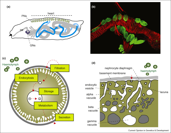
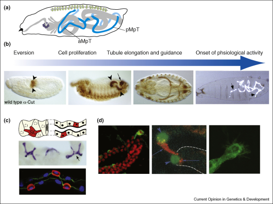
The function of all animal excretory systems is to rid the body of toxins and to maintain homeostatic balance. Although excretory organs in diverse animal species appear superficially different they are often built on two common principals: filtration and tubular secretion/reabsorbtion. The Drosophila excretory system is composed of filtration nephrocytes and Malpighian (renal) tubules. Here we review recent molecular genetic data on the development and differentiation of nephrocytes and renal tubules. We focus in particular on the molecular mechanisms that underpin key cell and tissue behaviours during morphogenesis, drawing parallels with other species where they exist. Finally we assess the implications of patterned tissue differentiation for the subsequent regulation of renal function. These studies highlight the continuing usefulness of the fly to provide fundamental insights into the complexities of organ formation.


Figures

Figure 1
Figure 1
(a) Cartoon to show the mature arrangement of pericardial (PNs, green circles) and garland (GNs, purple circles) nephrocytes, and Malpighian tubules (blue) in the late embryo/larva. The heart (linear grey structure) runs along the dorsal (upper) side of the animal. The alimentary canal (including oesophagus, oe) is depicted in grey. (b) Third instar larval pericardial nephrocytes (green, anti-Sns. Smaller stained cells, bottom are fat body) lined up on either side of the heart (red, phalloidin stain for actin. Alary muscles also appear red). Anterior to the left. (c) Cartoon of a nephrocyte. Haemolyph is filtered across basement membrane (grey outer circle) and nephrocyte diaphragm (black line linking nephrocyte foot processes) entering the lacunae, from where it is endocytosed (small white circle). Endocytosed material is stored and/or metabolised (large white circle). The stored and/or metabolised material can be secreted back into the haemolymph. Haemocytes (green circles). (d) Cartoon to show detail of the nephrocyte cortical region. Salient features including basement membrane, nephrocyte diaphragm and lacunae are labelled. Endocytosis from lacunae walls and foot process tips (red arrow) are shown. Vacuoles of different type (alpha, beta and gamma) are located within the cell. Haemocytes (green circles).
Figure 2
Figure 2
(a) Cartoon to show the mature arrangement of the nephrocytes (hollow circles) and Malpighian tubules (blue) in the larva (late embryo/larva). Anterior (aMpT) and posterior (pMpT) are labelled. (b) Sequence of developmental events underlying renal tubule maturation. Panels show embryos stained for Cut protein at stages 10, 12, 14 and shown in polarised light stage 17. Arrowheads show the developing tubules, the small arrow the posterior spiracle (also Cut-positive) and large arrows precipitates of uric acid in the mature tubule lumen. (c) Principal (no colour) and stellate (red in cartoon and lower panel, blue in central panel) cells in the distal, secretory region of adult (central) and 3rd instar larval tubules. As they integrate SCs become polarised (nuclei in blue, actin in green in lower panel), only developing a stellate morphology in the adult (arrow, central panel). (d) Tubule tip cells have a prominent morphology, secrete the EGF ligand sSpitz and show dynamic filipodial activity during tubule elongation. First panel stained for Cut (red) and CD8-GFP (green). Second panel ase-nLacZ line stained for β-Gal (green, arrowheads) and Rhomboid (red, arrows) showing both the tip cell and its sibling. Third panel CD8-GFP line showing the tip cell with many filopodial membrane extensions.

References

    1. Rafaeli-Bernstein A., Mordue W. The transport of the cardiac glycoside oubain by the Malpighian tubule of Zonocerus variegatus. Physiol Entomol. 2008;3:58–63.
    1. Vize P., Woolf A.S., Bard J.B.L. Academic Press; Orlando, FL: 2003. The Kidney, From Normal Development to Congenital Disease.
    1. McKanna J.A. Fine structure of the protonephridial system in Planaria. I. Flame cells. Z Zellforsch Mikrosk Anat. 1968;92:509–523. - PubMed
    1. Coons L.B., Alberti G. Acari, ticks. In: Harrison F.W., Foelix R.F., editors. vol 8B. Wiley–Liss; 1999. pp. 267–514. (Microscopic Anatomy of Invertebrates). Chelicerate Arthropoda.
    1. Farley RD: Scorpiones. In Microscopic Anatomy of Invertebrates, vol 8A. Edited by Harrison FW, Foelix RF. Wiley–Liss; 1999:117-222. Chelicerate Arthropoda.

Publication types

LinkOut - more resources