

doi: 10.1016/j.bmcl.2009.09.004.
Epub 2009 Sep 6.
Cellular localization and allele-selective inhibition of mutant huntingtin protein by peptide nucleic acid oligomers containing the fluorescent nucleobase [bis-o-(aminoethoxy)phenyl]pyrrolocytosine
Affiliations
- PMID: 19783436
- PMCID: PMC2770837
- DOI: 10.1016/j.bmcl.2009.09.004
Item in Clipboard
Cellular localization and allele-selective inhibition of mutant huntingtin protein by peptide nucleic acid oligomers containing the fluorescent nucleobase [bis-o-(aminoethoxy)phenyl]pyrrolocytosine
Bioorg Med Chem Lett.
.
Abstract
Peptide nucleic acid (PNA) is a successful DNA/RNA mimic. A major challenge for research is to invent chemically modified PNAs that retain the favorable properties of the parent compound while improving biological recognition. Here, we test modified PNAs containing [bis-o-(aminoethoxy)phenyl]pyrrolocytosine bases designed to engage guanine with an additional hydrogen bond. We observe elevated melting temperatures, localization to cellular compartments, and allele-selective inhibition of mutant huntingtin protein expression.
Figures
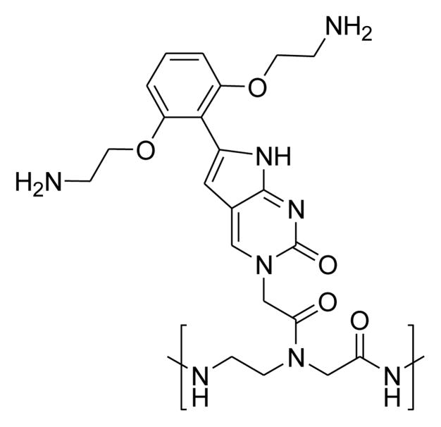
Structure of [bis-o-(aminoethoxy)phenyl]pyrrolocytosine.
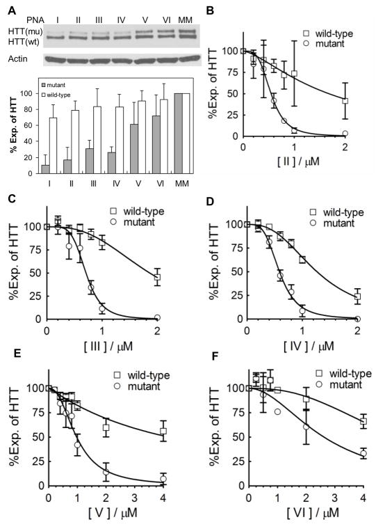
Modified PNAs selectively inhibit mutant (mu) relative to wild-type (wt) HTT expression in fibroblast cell line GM04281. A. Top, western analysis the effects of PNAs I–VI on HTT expression. Bottom, quantitation of inhibition of mutant and wild-type HTT by PNAs I–VI. PNAs were added at 1 μM concentration. B–F. Effects of PNAs II–VI on HTT expression at varied concentrations. Experiments were performed in triplicate. Expression is relative to expression to untreated cells.
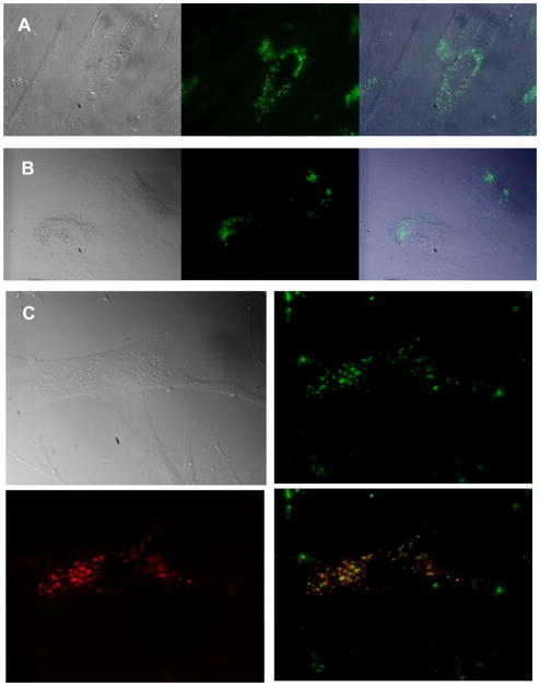
Fluorescent microscopy of PNA II in living fibroblasts. PNA was added at 1 μM concentration. A. One day or B. Nine days after PNA transfection. Left, Differential interference contrast microscopy (DIC) image; middle, PNA fluorescent; right, overlay of DIC and fluorescent images. C. PNA was co-localized with endosome marker Transferrin. 1 μM of Htt2 was co-incubated with 25 μg/mL of Transferrin-Alexa Fluor 633 for 15 h in fibroblast cells. Upper left, DIC image; upper right, PNA alone; lower left, transferrin fluorescence; lower right, overlay of PNA and transferrin images.
References
-
- Nielsen PE. Mol Biotech. 2004;26:233. - PubMed
-
- Nielsen PE, Egholm M, Berg RH, Buchardt O. Science. 1991;254:1497. - PubMed
-
- Egholm M, Buchardt O, Christensen L, Behrens C, Freier SM, Driver DA, Berg RH, Kim SK, Norden B, Nielsen PE. Nature. 1993;365:566. - PubMed
-
- Smulevitch SV, Simmons CG, Norton JC, Wise TW, Corey DR. Nat Biotech. 1996;14:1700. - PubMed
Publication types
MeSH terms
Substances
Grants and funding
LinkOut - more resources
Full Text Sources
Other Literature Sources
Miscellaneous
