. 2009 Dec 15;18(24):4843-52.
doi: 10.1093/hmg/ddp456. Epub 2009 Sep 25.

Preventing Ataxin-3 protein cleavage mitigates degeneration in a Drosophila model of SCA3

Affiliations

Preventing Ataxin-3 protein cleavage mitigates degeneration in a Drosophila model of SCA3

Joonil Jung et al. Hum Mol Genet. .

Abstract

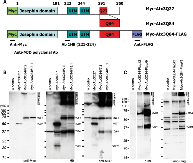
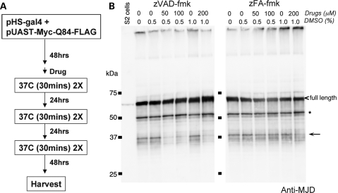
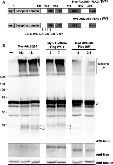
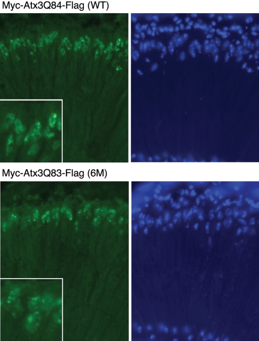
Protein cleavage is a common feature in human neurodegenerative disease. Ataxin-3 protein with an expanded polyglutamine (polyQ) repeat causes spinocerebellar ataxia type-3 (SCA3), also called Machado-Joseph disease, and is cleaved in mammalian cells, transgenic mice and SCA3 patient brain tissue. However, the pathological significance of Ataxin-3 cleavage has not been carefully examined. To gain insight into the significance of Ataxin-3 cleavage, we developed a Drosophila SL2 cell-based model as well as transgenic fly models. Our data indicate that Ataxin-3 protein cleavage is conserved in the fly and may be caspase-dependent as reported previously. Importantly, comparison of flies expressing either wild-type or caspase-site mutant proteins indicates that Ataxin-3 cleavage enhances neuronal loss in vivo. This genetic in vivo confirmation of the pathological role of Ataxin-3 cleavage indicates that therapies targeting Ataxin-3 cleavage might slow disease progression in SCA3 patients.


Figures

Figure 1.
Figure 1.
Ataxin-3 protein cleavage products are generated in Drosophila. (A) Schematic representation of Ataxin-3 proteins used. Site of the epitopes for the antibodies used are also indicated. (B) and (C) Western immunoblot analysis of proteins expressed in the brain of 7d fly heads. (B) Initial studies performed with Myc-Atx3Q27 and Myc-Atx3Q84 proteins. Left, Anti-Myc antibody detects the full-length proteins. Middle, the mouse monoclonal antibody 1H9 and right, the polyclonal anti-MJD antibody detect Ataxin-3 fragments migrating at ∼22 kDa for Atx3Q27 and ∼37 kDa for Atx3Q84. Each antibody also detects non-specific bands when compared with the control. Strong signals are detected as SDS-insoluble fractions in the stacking gel. (C) Left, transgenic Drosophila head extracts expressing Myc-Atx3Q84-Flag show a similar cleavage pattern by 1H9, of the full-length protein and ∼37 kDa fragments. Right, anti-Flag antibody detects the full-length protein and a ∼37 kDa fragment. These data suggest that ∼37 kDa fragments, also reported in transgenic mouse and some MJD patient samples, are likely to contain the C-terminal portion of MJD protein, as well as the polyQ domain of the protein. Genotypes: (B) elav-gal4/+; UAS-Myc-Atx3Q27/+ or UAS-Myc-Atx3Q84/+ and (C) elav-gal4/+; UAS-Myc-Atx3Q84-Flag/+. Full-length proteins, <Q84 or <Q27, fragments indicated as *Q27f and *Q84f.
Figure 2.
Figure 2.
zVAD, a broad-acting caspase inhibitor, can suppress Ataxin-3 protein cleavage in an SL2-cell-based system. (A) Experimental procedure to detect the effect of various protease inhibitors on Ataxin-3 protein cleavage in SL2 cells. Expression of the Ataxin-3 protein was driven by heat shock using a Gal4 (pHS-gal4) construct. The construct in all cases was pUAST-Myc-Atx3Q84. Drug was added 1 h prior to the first heat shock and was then continuously present until harvesting of the cells. (B) Ataxin-3 protein cleavage pattern in SL2 cells. The full-length protein is marked with a carrot (<) and the ∼37 kDa fragments highlighted with an arrow. In addition to the ∼37 kDa fragments seen in the fly in vivo, additional Ataxin-3 fragments were detected in SL2 cells, most prominently a fragments at ∼50 kDa (*). zVAD, a general caspase inhibitor, inhibited the production of the ∼37 kDa fragments, without significantly altering the generation of the ∼50 kDa fragment. zFA, a control peptide, had no effect. All drugs were dissolved in DMSO, with final percentage of DMSO indicated.
Figure 3.
Figure 3.
Mutation of putative caspase recognition sites in Ataxin-3 dramatically reduces the amount of cleavage product in vivo. (A) Schematic diagram of Atx3Q83 protein with the positions noted of the six caspase sites targeted in the site-directed mutagenesis (D->N). (B) Western immunoblots. Top panel, Ataxin-3 protein cleavage pattern probed with anti-MJD polyclonal antibody. Head extracts from 7d adult flies were used. Sextuplet mutation strongly suppresses Ataxin-3 protein cleavage. A non-specific band (*, ∼70 kDa) masks the full-length band. SDS-insoluble fractions are within the stacking gel, and the ∼37 kDa cleavage products (**U and **D) are marked. Genotypes: elav-gal4/UAS-Myc-Atx3Q84, elav-gal4/ UAS-Myc-Atx3Q84-Flag (WT) and elav-gal4/UAS-Myc-Atx3Q83-Flag (6M). Two transgenic lines of each genotype are shown. Lower panels are the same blot, probed with Anti-Myc for the full-length protein, and with Anti-tubulin, for a loading control.
Figure 4.
Figure 4.
Elimination of putative caspase cleavage sites in Ataxin-3 protein has little impact on protein accumulations. Cryosections immunostained for Ataxin-3 protein with Myc (green, left panels) and for nuclei with Hoechst (blue, right panels) of retinal sections from 7d adult flies. Protein was expressed with an adult onset driver (rh1-gal4), which is a sensitive method to detect subtle differences in nuclear inclusion formation. Flies expressing either the WT protein (top) or the 6M protein (bottom) showed inclusions typical of the full-length Ataxin-3 protein, being of irregular shape when assayed with this late-onset driver, and had the same onset and size. Higher magnification insets are also included in the Myc panels. Genotypes: rh1-gal4/ UAS-Myc-Atx3Q84-Flag (WT) and rh1-gal4/ UAS-Myc-Atx3Q83-Flag (6M).
Figure 5.
Figure 5.
Suppression of Ataxin-3 protein cleavage slows down the progression of photoreceptor neuronal degeneration. (A) Images of the retina by pseudopupil technique, showing 1d and 15d representative images of flies expressing Myc-Atx3Q84-Flag (WT) and Myc-Atx3Q83-Flag (6M). (B) Quantitation of the number of photoreceptor neurons in 1d WT (green) and 6M (red) flies. At 1d, flies of both genotypes showed a similar degree of mild photoreceptor neuron degeneration. Average number of photoreceptor neurons indicated ± SD. 15d adult animals show greater degeneration in flies expressing the WT protein than the 6M protein. Genotypes: WT, elav-gal4/+; UAS-Myc-Atx3Q84-Flag (WT)/+ and 6M, elav-gal4/+; pUAS-Myc-Atx3Q83-Flag (6M)/+. (C) The 6M protein retains normal Ataxin-3 activity to suppress pathogenicity of the truncated Q78 protein. External eye pictures. Flies expressing the truncated Q78 protein alone (—) have a degenerate eye. The normal non-pathogenic Ataxin-3 protein [WT(Q27)] suppresses this toxicity when co-expressed; this suppression reflects the normal functional activity of the Ataxin-3 protein (13). The pathogenic protein (WT) also shows suppressor activity, although not as striking as the non-pathogenic protein. The 6M protein (6M) has suppressor activity similar to the WT pathogenic protein. This indicates that the 6M protein is likely properly folded because it retains normal activity of the Ataxin-3 protein to suppress polyQ toxicity. Genotypes: gmr-gal4 UAS-SCA3trQ78(S)/+. gmr-gal4 UAS-SCA3trQ78(S)/UAS-Myc-Atx3Q27-Flag [WT(Q27)]. gmr-gal4 UAS-SCA3trQ78(S)/UAS-Myc-Atx3Q84-Flag (WT). gmr-gal4 UAS-SCA3trQ78(S)/ UAS-Myc-Atx3Q83-Flag (6M).

References

    1. Beher D. Gamma-secretase modulation and its promise for Alzheimer's disease: a rationale for drug discovery. Curr. Top. Med. Chem. 2008;8:34–37. - PubMed
    1. Rohn T.T., Head E. Caspases as therapeutic targets in Alzheimer's disease: is it time to ‘cut’ to the chase? Int. J. Clin. Exp. Pathol. 2009;2:108–118. - PMC - PubMed
    1. Gamblin T.C., Chen F., Zambrano A., Abraha A., Lagalwar S., Guillozet A.L., Lu M., Fu Y., Garcia-Sierra F., LaPointe N., et al. Caspase cleavage of tau: linking amyloid and neurofibrillary tangles in Alzheimer's disease. Proc. Natl Acad. Sci. USA. 2003;100:10032–10037. - PMC - PubMed
    1. Newman J., Rissman R.A., Sarsoza F., Kim R.C., Dick M., Bennett D.A., Cotman C.W., Rohn T.T., Head E. Caspase-cleaved tau accumulation in neurodegenerative diseases associated with tau and alpha-synuclein pathology. Acta Neuropathol. 2005;110:135–144. - PubMed
    1. Hoffner G., Island M.L., Djian P. Purification of neuronal inclusions of patients with Huntington's disease reveals a broad range of N-terminal fragments of expanded huntingtin and insoluble polymers. J. Neurochem. 2005;95:125–136. - PubMed

Publication types

MeSH terms