. 2009 Dec;191(23):7234-42.
doi: 10.1128/JB.00923-09. Epub 2009 Sep 25.

Distribution and phylogeny of light-oxygen-voltage-blue-light-signaling proteins in the three kingdoms of life

Affiliations

Distribution and phylogeny of light-oxygen-voltage-blue-light-signaling proteins in the three kingdoms of life

Ulrich Krauss et al. J Bacteriol. 2009 Dec.

Abstract

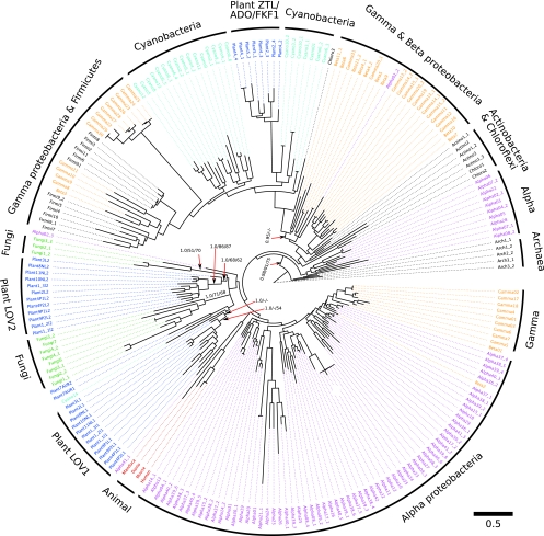
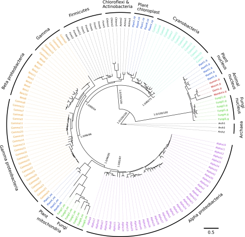
Plants and fungi respond to environmental light stimuli via the action of different photoreceptor modules. One such class, responding to the blue region of light, is constituted by photoreceptors containing so-called light-oxygen-voltage (LOV) domains as sensor modules. Four major LOV families are currently identified in eukaryotes: (i) the plant phototropins, regulating various physiological effects such as phototropism, chloroplast relocation, and stomatal opening; (ii) the aureochromes, mediating photomorphogenesis in photosynthetic stramenopile algae; (iii) the plant circadian photoreceptors of the zeitlupe (ZTL)/adagio (ADO)/flavin-binding Kelch repeat F-box protein 1 (FKF1) family; and (iv) the fungal circadian photoreceptors white-collar 1 (WC-1). Blue-light-sensitive LOV signaling modules are also widespread throughout the prokaryotic world, and physiological responses mediated by bacterial LOV photoreceptors were recently reported. Thus, the question arises as to the evolutionary relationship between the pro- and eukaryotic LOV photoreceptor systems. We used Bayesian and maximum-likelihood tree reconstruction methods to infer evolutionary scenarios that might have led to the widespread appearance of LOV domains among the pro- and eukaryotes. The phylogenetic study presented here suggests a bacterial origin for the LOV domains of the four major eukaryotic LOV photoreceptor families, whereas the LOV sensor domains were most likely recruited from the bacteria in the course of plastid and mitochondrial endosymbiosis.


Figures

FIG. 1.
FIG. 1.
Phylogenetic tree determined by RAxML rapid bootstrap (55), reconstructed for the LOV gene sequences from divergent taxa. In addition, Bayesian analysis was performed using MrBayes (24) and bootstrap with ML by IQPNNI (62a) as described in Materials and Methods. Bayesian posterior probabilities (BPP) and RAxML and IQPNNI (in this order) bootstrap support values (BP) are shown at the relevant branches of the RAxML bootstrap consensus tree. The respective LOV sequences of the different eukaryotic photoreceptor families as well as the LOV sequences of prokaryotic origin are color coded: the Archaea (outgroup), the Actinobacteria, Chloroflexi, and Firmicutes, are shown in black, Alphaproteobacteria are in pink, Betaproteobacteria and Gammaproteobacteria are in orange, cyanobacterial genera are in cyan, fungal WC-1 sequences are in green, plant phototropin and neochrome LOV domains (LOV1 and LOV2), algal aureochrome LOV sequences (Plant7AUR2 and Plant7AUR1), and the ZTL/ADO/FKF1-LOV family are in blue, and the putative animal LOV domains are in red. Detailed sequence information, including protein accession numbers, is summarized in Table S1 in the supplemental material.
FIG. 2.
FIG. 2.
SSU rRNA gene tree reconstructed to infer the species evolution of the LOV domain-containing taxa. Bayesian, bootstrap, and ML trees were constructed as described for the LOV gene tree. All trees that were generated using the different phylogeny inference methods showed the same overall topology. Here, the MrBayes tree is depicted. Bayesian posterior probabilities (BPP) and bootstrap support values (BP) with RAxML and IQPNNI are added at relevant branches. The color code is the same as in Fig. 1. The suffixes g, mt, and cp indicate SSU rRNA sequences from nuclear, mitochondrial, and chloroplast regions, respectively (see Table S1 in the supplemental material for details). The branch lengths of the subtree containing mitochondrial rRNA were scaled down three times to better fit in the figure.

References

    1. Altschul, S. F., W. Gish, W. Miller, E. W. Myers, and D. J. Lipman. 1990. Basic local alignment search tool. J. Mol. Biol. 215:403-410. - PubMed
    1. Altschul, S. F., T. L. Madden, A. A. Schaffer, J. Zhang, Z. Zhang, W. Miller, and D. J. Lipman. 1997. Gapped BLAST and PSI-BLAST: a new generation of protein database search programs. Nucleic Acids Res. 25:3389-3402. - PMC - PubMed
    1. Ballario, P., P. Vittorioso, A. Magrelli, C. Talora, A. Cabibbo, and G. Macino. 1996. White collar-1, a central regulator of blue light responses in Neurospora, is a zinc finger protein. EMBO J. 15:1650-1657. - PMC - PubMed
    1. Battistuzzi, F. U., A. Feijao, and S. B. Hedges. 2004. A genomic timescale of prokaryote evolution: insights into the origin of methanogenesis, phototrophy, and the colonization of land. BMC Evol. Biol. 4:44. - PMC - PubMed
    1. Boer, P. H., and M. W. Gray. 1988. Scrambled ribosomal RNA gene pieces in Chlamydomonas reinhardtii mitochondrial DNA. Cell 55:399-411. - PubMed

Publication types

MeSH terms

LinkOut - more resources