. 2009 Nov 20;284(47):32782-91.
doi: 10.1074/jbc.M109.054528. Epub 2009 Sep 25.

Involvement of the beta clamp in methyl-directed mismatch repair in vitro

Affiliations

Involvement of the beta clamp in methyl-directed mismatch repair in vitro

Anna Pluciennik et al. J Biol Chem. .

Abstract

We have examined function of the bacterial beta replication clamp in the different steps of methyl-directed DNA mismatch repair. The mismatch-, MutS-, and MutL-dependent activation of MutH is unaffected by the presence or orientation of loaded beta clamp on either 3' or 5' heteroduplexes. Similarly, beta is not required for 3' or 5' mismatch-provoked excision when scored in the presence of gamma complex or in the presence of gamma complex and DNA polymerase III core components. However, mismatch repair does not occur in the absence of beta, an effect we attribute to a requirement for the clamp in the repair DNA synthesis step of the reaction. We have confirmed previous findings that beta clamp interacts specifically with MutS and MutL (López de Saro, F. J., Marinus, M. G., Modrich, P., and O'Donnell, M. (2006) J. Biol. Chem. 281, 14340-14349) and show that the mutator phenotype conferred by amino acid substitution within the MutS N-terminal beta-interaction motif is the probable result of instability coupled with reduced activity in multiple steps of the repair reaction. In addition, we have found that the DNA polymerase III alpha catalytic subunit interacts strongly and specifically with both MutS and MutL. Because interactions of polymerase III holoenzyme components with MutS and MutL appear to be of limited import during the initiation and excision steps of mismatch correction, we suggest that their significance might lie in the control of replication fork events in response to the sensing of DNA lesions by the repair system.


Figures

FIGURE 1.
FIGURE 1.
Assembly of β clamp in the presence of γ complex onto supercoiled or nicked DNA. A, 3′ nicked G-T heteroduplex (lanes 1 and 2) or 3′ G-T supercoiled heteroduplex (lanes 3 and 4) were incubated with 32P-labeled β clamp and γ complex in the absence (lanes 1 and 3) or presence of 30 mm NaCl (lanes 2 and 4), β·DNA complexes cross-linked, and the products were resolved by electrophoresis through 1% agarose (“Experimental Procedures”). DNA was visualized after ethidium staining (left panel) and β·DNA complexes scored by phosphorimaging (right panel). The positions of the nicked open circular (oc) DNA, supercoiled (sc) DNA, and free β are shown on the right. The stoichiometry of β loading was determined from percentage of the total signal in each lane. B, lifetime of β·DNA complexes on supercoiled G-T heteroduplex DNA was determined by loading [32P]β in the absence of NaCl, followed by the addition of NaCl to 100 mm to prevent further loading. Incubation was continued, and the samples were removed and scored for β·DNA complexes (“Experimental Procedures”). Lanes 1–5, samples removed 0, 5, 10, 20, and 30 min after 100 mm NaCl addition. Lanes 6–10, assembly reaction was performed in the presence of 100 mm NaCl, and the samples were taken at 0, 5, 10, 20, and 30 min. β·DNA complexes were visualized as in A.
FIGURE 2.
FIGURE 2.
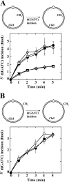
Loaded β clamp is not required for MutH activation. A, diagram depicts incision of a 3′ G-T supercoiled hemimethylated heteroduplex by activated MutH. Substrates contained a G-T mismatch (A·T base pair in homoduplex control) and d(GATC) methylation on the complementary DNA strand (unmethylated d(GATC) sequence 3′ to mismatch as viewed along the shorter path between the two sites). The 3′ G-T heteroduplex was preincubated with 150 nm β clamp and 12 nm γ complex (open circles), preincubated with 300 nm β clamp and 16 nm γ complex (diamonds), or mock preincubated (β clamp and γ complex omitted) (closed circles) at low ionic strength to ensure a proper assembly of β clamp onto DNA (“Experimental Procedures”). Control A·T homoduplex was preincubated with 300 nm β and 16 nm γ complex (open triangles) or mock preincubated (inverted closed triangles) in a similar manner. The products were used immediately for MutH activation assays (“Experimental Procedures”). The error bars represent one standard deviation for three independent experiments. B, experimental procedures and symbols are as in A except the substrate was a 5′ G-T supercoiled hemimethylated heteroduplex with d(GATC) modification on the viral DNA strand.
FIGURE 3.
FIGURE 3.
Orientation of β loading does not affect MutH activation. Open circular G-T heteroduplex (or A·T homoduplex) DNAs contained a single strand break within the complementary DNA strand 128 bp 3′ to the mismatch (shorter path), as well as d(GATC) methylation on the complementary DNA strand (A, 3′ heteroduplex/homoduplex) or viral strand (B, 5′ heteroduplex/homoduplex). DNAs were preincubated with β clamp and γ complex (G-T, open circles; A·T, open triangles) or mock preincubated (G-T, closed circles; A·T, closed inverted triangles) as described under “Experimental Procedures.” The products were used immediately for MutH activation assays. The error bars represent one standard deviation for three independent experiments.
FIGURE 4.
FIGURE 4.
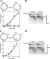
β clamp is essential for the repair synthesis step of mismatch correction. Supercoiled 3′ (A and B; dGATC methylation on complementary strand) or 5′ (C and D; methylation on viral strand) G-T heteroduplexes were incubated with MutS, MutL, MutH, DNA helicase II, SSB, Pol III core, γ complex, dNTPs, and exonuclease I (3′ heteroduplex) or RecJ (5′ heteroduplex) in the presence or absence of β clamp (“Experimental Procedures”). The G-T mismatch in substrate DNAs resides within overlapping HindIII and XhoI sites, rendering the heteroduplexes resistant to both enzymes (30). Repair of 3′ (A) or 5′ (C) heteroduplexes confers XhoI or HindIII sensitivity, respectively (closed circles, + β clamp; open circles, β omitted from reactions). The error bars correspond to one standard deviation for three independent measurements. The reaction products were also digested with ClaI and analyzed by indirect end labeling to visualize excision repair end points produced during the reaction (“Experimental Procedures”) as illustrated in the diagrams on the right. Excision on the unmethylated DNA strand proceeds 3′ to 5′ (3′ heteroduplex) or 5′ to 3′ (5′ heteroduplex) toward the mismatch from the MutH-produced strand break at the d(GATC) site (33). The arrows adjacent to the diagrams indicate the direction of DNA synthesis on the repaired strand. The asterisk in D indicates a DNA terminus that was present in a subset of 5′ heteroduplex molecules because of incomplete ligation of a strand break during substrate preparation.
FIGURE 5.
FIGURE 5.
β clamp does not influence the excision step of methyl-directed mismatch repair. Supercoiled 3′ (A and B; dGATC methylation on complementary strand) or 5′ (C and D; methylation on viral strand) G-T heteroduplexes were preincubated with β clamp, γ complex, and Pol III core (A and C, open circles) or mock preincubated (A and C, closed circles). Supercoiled 3′ A·T homoduplex control DNA was also subjected to mock preincubation (A, inverted triangles). The products were used immediately for mismatch-provoked excision assays, which contained MutS, MutL, DNA helicase II, SSB, dATP, dGTP, dCTP, ddTTP, and exonuclease I (3′ heteroduplex/homoduplex) or RecJ exonuclease (5′ heteroduplex). Substrates contain an NheI site 5 bp from the location of the mispair, which is rendered resistant to cleavage by mismatch-provoked excision (“Experimental Procedures”). Extents of excision scored by this assay are shown in A and C. Heteroduplex reaction products were also digested with ClaI and analyzed by indirect end labeling to visualize excision end points produced in the absence or presence of Pol III components (B and D). The asterisk in D indicates a DNA terminus that was present in a subset of 5′ heteroduplex molecules because of incomplete ligation of a strand break during substrate preparation.
FIGURE 6.
FIGURE 6.
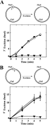
β clamp does not influence the nick-directed mismatch excision. Open circular G-T heteroduplex DNAs (closed circles, open circles, and open diamonds), or control A·T homoduplexes (inverted triangles) contained a single-strand break in the complementary strand 128 bp 3′ (A) or 128 bp 5′ (B) to the location of the mismatch. Nicked DNAs were preincubated with β clamp and γ complex (open circles); preincubated with β clamp, γ complex and Pol III core (open diamonds); or mock preincubated (closed circles and inverted triangles). Products were used immediately for mismatch-provoked excision assays, which contained MutS, MutL, DNA helicase II, SSB, dATP, dGTP, dCTP, ddTTP, and exonuclease I (3′ strand break) or RecJ exonuclease (5′ strand break). Excision was scored by NheI resistance assay (Fig. 5 and “Experimental Procedures”). The error bars for the heteroduplex with a 5′ strand break correspond to one standard deviation for three independent experiments. The results shown for the heteroduplex with a 3′ strand break are the averages of two determinations.
FIGURE 7.
FIGURE 7.
Activity comparisons for wild type MutS or MutSN. A, supercoiled 3′ G-T hemimethylated heteroduplex (Fig. 2A) was incubated with MutH, MutL, and variable concentrations of wild type MutS (closed circles) or MutSN (open circles) in the absence of Pol III components. The reactions were sampled as a function of time and initial rates of d(GATC) incision determined (“Experimental Procedures”). B, supercoiled 3′ G-T hemimethylated heteroduplex was incubated with MutH, MutL, DNA helicase II, SSB, exonuclease I, and variable concentrations of wild type MutS (closed circles) or MutSN (open circles) under excision conditions (“Experimental Procedures”) except that Pol III components, ddTTP, and dNTPs were omitted. The reactions were sampled as a function of time to determine initial rates of excision, which were quantified by NheI resistance assay (Fig. 5). C, supercoiled 3′ G-T hemimethylated heteroduplex was incubated under repair conditions (“Experimental Procedures”) with MutL, MutH, DNA helicase II, SSB, exonuclease I, Pol III core, β clamp, γ complex, and MutS (closed circles) or MutSN (open circles) as indicated. The samples were taken as a function of time and repair quantified by XhoI-sensitivity assay (Fig. 4A) to determine initial rates of repair. D, extracts of E. coli BT199ΔmutS2K6 (harboring pET-mutSwt or pET-mutSN) were analyzed for MutS protein by Western blot as described under “Experimental Procedures.” Ponceau S staining of the Western membrane was used as a loading control.
FIGURE 8.
FIGURE 8.
Interactions of MutL and MutS with components of Pol III holoenzyme. β clamp, γ complex, Pol III core, BSA, MutS, MutL, and DNA helicase II were applied as indicated to nitrocellulose membranes (upper panels). Alternatively, proteins samples were subjected to SDS-PAGE on 10% gels (lower panels; 4-pmol sample load) followed by transfer to nitrocellulose membranes. The membranes were used for far Western analysis by incubation with MutL (A) or MutS (B), followed by immunochemical visualization of membrane-bound MutL and MutS (“Experimental Procedures”). SDS gel species that bind MutL and MutS are indicated to the right of each gel transfer, with identification based on parallel gels that were stained with Coomassie Blue. No signals were observed with otherwise identical membranes when MutS or MutL incubation steps were omitted (not shown).

Similar articles

Cited by

References

    1. Iyer R. R., Pluciennik A., Burdett V., Modrich P. L. (2006) Chem. Rev. 106, 302–323 - PubMed
    1. Li L. S., Morales J. C., Hwang A., Wagner M. W., Boothman D. A. (2008) J. Biol. Chem. 283, 21394–21403 - PMC - PubMed
    1. Hsieh P., Yamane K. (2008) Mech. Ageing Dev. 129, 391–407 - PMC - PubMed
    1. Pukkila P. J., Peterson J., Herman G., Modrich P., Meselson M. (1983) Genetics 104, 571–582 - PMC - PubMed
    1. Lahue R. S., Au K. G., Modrich P. (1989) Science 245, 160–164 - PubMed

Publication types

MeSH terms

LinkOut - more resources