. 2009 Nov 20;284(47):32959-67.
doi: 10.1074/jbc.M109.036129. Epub 2009 Sep 25.

ScFv antibody-induced translocation of cell-surface heparan sulfate proteoglycan to endocytic vesicles: evidence for heparan sulfate epitope specificity and role of both syndecan and glypican

Affiliations

ScFv antibody-induced translocation of cell-surface heparan sulfate proteoglycan to endocytic vesicles: evidence for heparan sulfate epitope specificity and role of both syndecan and glypican

Anders Wittrup et al. J Biol Chem. .

Abstract

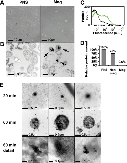
Cellular uptake of several viruses and polybasic macromolecules requires the expression of cell-surface heparan sulfate proteoglycan (HSPG) through as yet ill defined mechanisms. We unexpectedly found that among several cell-surface-binding single chain variable fragment (scFv) anti-HS antibody (alphaHS) clones, only one, AO4B08, efficiently translocated macromolecular cargo to intracellular vesicles through induction of HSPG endocytosis. Interestingly, AO4B08-induced PG internalization was strictly dependent on HS 2-O-sulfation and appeared independent of intact N-sulfation. AO4B08 and human immunodeficiency virus (HIV)-Tat, i.e. a well known cell-penetrating peptide, were shown to compete for the internalizing PG population. To obtain a more detailed characterization of this pathway, we have developed a procedure for the isolation of endocytic vesicles by conjugating AO4B08 with superparamagnetic nanoparticles. [(35)S]sulfate-labeled HSPG was found to accumulate in isolated, AO4B08-containing vesicles, providing the first biochemical evidence for intact HSPG co-internalization with its ligand. Further analysis revealed the existence of both syndecan, i.e. a transmembrane HSPG, and glycosyl-phosphatidyl-inositol-anchored glypican in purified vesicles. Importantly, internalized syndecan and glypican were found to co-localize in AO4B08-containing vesicles. Our data establish HSPGs as true internalizing receptors of macromolecular cargo and indicate that the sorting of cell-surface HSPG to endocytic vesicles is determined by a specific HS epitope that can be carried by both syndecan and glypican core protein.


Figures

FIGURE 1.
FIGURE 1.
Epitope-specific αHS internalization. HeLa (A) and A549 (B) cells were surface-stained at 4 °C with vsv-tagged αHS followed by mouse anti-vsv and Alexa Fluor 488-conjugated anti-mouse antibodies (gray bars). Similar experiments were performed at 37 °C to allow for antibody internalization (black bars), showing substantial internalization of clone AO4B08. Antibody complex binding and uptake were analyzed by flow cytometry as described under “Experimental Procedures.” C and D, αHS antibody uptake is through endocytosis. HeLa cells were incubated for the indicated times with AO4B08-F (red), and nuclei were counterstained with ToPro (blue) and visualized using confocal microscopy. D, HeLa cells were incubated for the indicated times with magnetic nanoparticle (100 nm) conjugated AO4B08 antibody complexes (AO4B08-M) and visualized using electron microscopy. Shown are representative pictures from at least three independent experiments. E, αHS uptake is strictly HS-dependent. Upper panels, uptake of AO4B08-F in HeLa cells following either no treatment (left panel) or pretreatment with chondroitinase ABC lyase (ABC'ase) or heparanase I and III lyases (HS'ase) to digest chondroitin/dermatan sulfate PG and HSPG, respectively. Lower panels, uptake of AO4B08-F in wild-type CHO-K1, pan PG-deficient pgsA-745, and HSPG-deficient pgsD-677 CHO cells as determined by flow cytometry. Filled curves, control cells incubated with mouse anti-vsv and fluorophore-labeled anti-mouse antibodies only. a. u., arbitrary units. F, AO4B08-F uptake requires 2-OS HSPG. Uptake of AO4B08-F or RB4EA12-F in wild-type (CHO-K1; light gray bars), 2-OS-deficient pgsF-17 (2-OS-def.; dark gray bars), and NS-deficient pgsE-606 (NS-def.; black bars) CHO cells as determined by flow cytometry. Data are presented as the mean S.D. (error bars).
FIGURE 2.
FIGURE 2.
AO4B08 and HIV-Tat compete for HS binding, and both induce internalization of cell-surface PGs. A, cell-surface binding of Tat-Alexa Fluor 647 (2 μg/ml) in HeLa cells in the presence of increasing concentrations of AO4B08 antibody (0–0.2, v/v). B, uptake of Tat-Alexa Fluor 647 (2 μg/ml) over a period of 30 min in the presence of increasing concentrations of AO4B08 antibody (0–0.2, v/v). C, AO4B08-F and DNA/Tat complexes co-localize in endocytic vesicles. Confocal microscopy analysis of HeLa cells co-incubated for 30 min with DNA-YOYO-1/Tat complexes (10 and 40 μg/ml, respectively; green, DNA-YOYO-1) and AO4B08-F (red) is shown. D, HeLa cells were incubated with various internalizing HS ligands, i.e. AO4B08 antibody complexes (αHS), Tat peptide alone, or DNA/Tat complexes for the indicated times at 37 °C. After washing with 1 m NaCl to remove the remaining surface-bound ligands, the amount of residual cell-surface HSPG was determined by surface stain with AO4B08 antibody at 4 °C followed by flow cytometry analysis. E, HeLa cells were incubated with DNA/Tat complexes or myc-tagged AO4B08 antibody complexes (αHS) for 180 min at 37 °C. After washing with 1 m NaCl, the amount of residual cell-surface AO4B08 and RB4EA12 epitope was determined by surface stain with vsv-tagged antibodies at 4 °C followed by flow cytometry analysis. Error bars represent S.D. F, cells were co-incubated for 30 min at 37 °C with myc-tagged AO4B08 antibody complexes (red) and vsv-tagged RB4EA12 antibody complexes (green) and visualized using confocal microscopy. Detail, magnification of the indicated area in panels to the left.
FIGURE 3.
FIGURE 3.
Isolation of endocytic vesicles containing magnetic nanoparticle-conjugated AO4B08 antibody complexes. A–E, HeLa cells were incubated with AO4B08-F and AO4B08-M for 1 h, extensively washed and trypsinized, and mechanically disrupted. The resulting subcellular particles were visualized using epifluorescence microscopy and differential interference contrast (A) or by electron microscopy (B) prior to (PNS) and after (Mag) magnetic separation. The arrows in B indicate vesicles containing magnetic particles. C, flow cytometry analysis of PNS (black curve) and magnetic (green curve) fractions shows the enrichment of fluorescent particles in the magnetic fraction. a. u., arbitrary units. D, relative protein amounts in the respective fraction from a representative experiment. Non-mag, non-magnetic. E, subcellular structures of the magnetic fraction were negatively stained and analyzed by electron microscopy, showing intact vesicles containing magnetic nanoparticles. Lower panel, high magnification images of the indicated areas show the dotted structure of magnetic particles.
FIGURE 4.
FIGURE 4.
Enrichment of high molecular weight PGs in magnetically isolated vesicles. A–C, HeLa cells were metabolically labeled with [35S]sulfate for ∼24 h and then incubated for 20, 40, 60, or 180 min with AO4B08-M. At the end of antibody incubation, cells were subjected to trypsinization, mechanical disruption, and magnetic separation. [35S]PG was isolated from the PNS, which was separated into the non-magnetic (Non-mag) and magnetic (Mag) fractions of PNS as described under “Experimental Procedures.” A, SDS-PAGE and autoradiography of isolated [35S]PG from the magnetic fraction shows time-dependent increase of intact, high molecular weight PGs. B, [35S]PG in the respective fraction per μg of total protein at the 60-min time point. C, [35S]PG from the respective fraction was separated by size-exclusion chromatography on a Superose 12 column. Data shown are representative of at least three independent experiments.
FIGURE 5.
FIGURE 5.
Magnetically isolated vesicles contain both SDC and GPC PGs strictly substituted with HS chains. A, HeLa cells were [35S]sulfate-labeled and incubated with AO4B08-M for 1 h as described in the legend for Fig. 4. Isolated [35S]PGs from the PNS (P) and the non-magnetic (N) and magnetic (M) fraction of PNS were either untreated (Control) or digested with heparanase I and III lyases (HS'ase) or chondroitinase ABC lyase (ABC'ase) followed by SDS-PAGE analysis. B, densitometric quantification of high molecular mass (>64 kDa) HSPGs in the respective fraction following extensive ABC lyase digestion. Data are presented as the relative amount of [35S]HSPG/μg of total protein. Mag, magnetic; Non-mag, non-magnetic. C, isolated PGs from a whole cell lysate of non-trypsinized control cells (cell lysate) or the non-magnetic and the magnetic fraction of the PNS from cells incubated with AO4B08-M as in A were digested with heparanase III and ABC lyases. The digest products were separated by SDS-PAGE, and HSPG core proteins were visualized by immunoblotting with 3G10 anti-ΔHS antibody. Non-digested PGs (second column from the left) showed virtually no signal, confirming the specificity of the 3G10 antibody. Arrow 1, position of GPC1 (see also panel E); arrow 2, position of SDC2 (see also panel E). D, quantitative real-time PCR analysis of HSPG core protein mRNA expression in HeLa cells. Error bars represent S.D. E, immunoblotting with 3G10 anti-ΔHS antibody and core protein-specific antibodies on purified total PG from cell extracts. F, immunoblotting with anti-GPC1 or anti-SDC2 antibodies, as indicated, of intact HSPGs purified from the magnetic fraction.
FIGURE 6.
FIGURE 6.
Both SDC and GPC mediate AO4B08 antibody complex internalization and can be distributed to the same subset of endocytic vesicles. A, HeLa cells transfected with SDC2-GFP, SDC3-GFP, or HA-GPC3 (green), as indicated, were incubated for 30 min with AO4B08 antibody complexes (red), as described under “Experimental Procedures.” Detail, merged images of the indicated areas show significant co-localization of AO4B08-F with all three HSPGs. B, Cells co-transfected with SDC2-GFP (green) and HA-GPC3 (red) were incubated for 10 min at 37 °C with AO4B08 antibody complexes (blue) and visualized using confocal microscopy. Under Detail, the arrows indicate co-association of SDC2-GFP, HA-GPC3, and AO4B08-F in vesicular structures close to the plasma membrane.

Similar articles

Cited by

References

    1. Mislick K. A., Baldeschwieler J. D. (1996) Proc. Natl. Acad. Sci. U.S.A. 93, 12349–12354 - PMC - PubMed
    1. Belting M., Persson S., Fransson L. A. (1999) Biochem. J. 338, 317–323 - PMC - PubMed
    1. Sandgren S., Cheng F., Belting M. (2002) J. Biol. Chem. 277, 38877–38883 - PubMed
    1. Sandgren S., Wittrup A., Cheng F., Jönsson M., Eklund E., Busch S., Belting M. (2004) J. Biol. Chem. 279, 17951–17956 - PubMed
    1. Payne C. K., Jones S. A., Chen C., Zhuang X. (2007) Traffic 8, 389–401 - PMC - PubMed

Publication types

LinkOut - more resources