. 2009 Oct 15;183(8):5146-57.
doi: 10.4049/jimmunol.0802610. Epub 2009 Sep 25.

Genetic evidence that the differential expression of the ligand-independent isoform of CTLA-4 is the molecular basis of the Idd5.1 type 1 diabetes region in nonobese diabetic mice

Affiliations

Genetic evidence that the differential expression of the ligand-independent isoform of CTLA-4 is the molecular basis of the Idd5.1 type 1 diabetes region in nonobese diabetic mice

Manabu Araki et al. J Immunol. .

Abstract

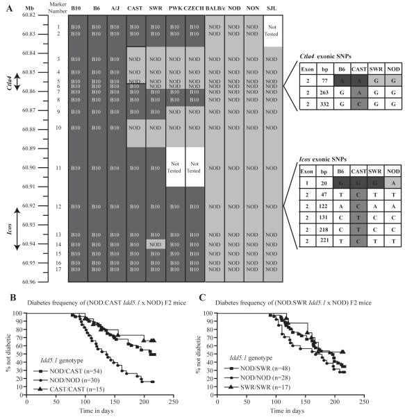
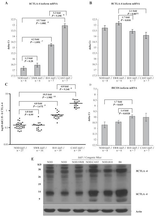
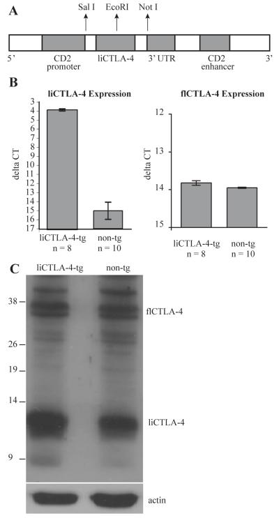
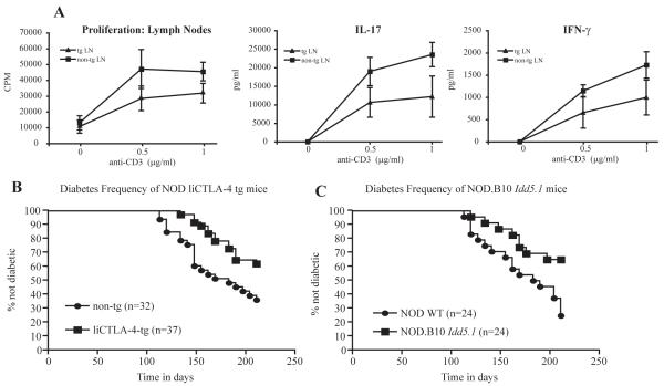
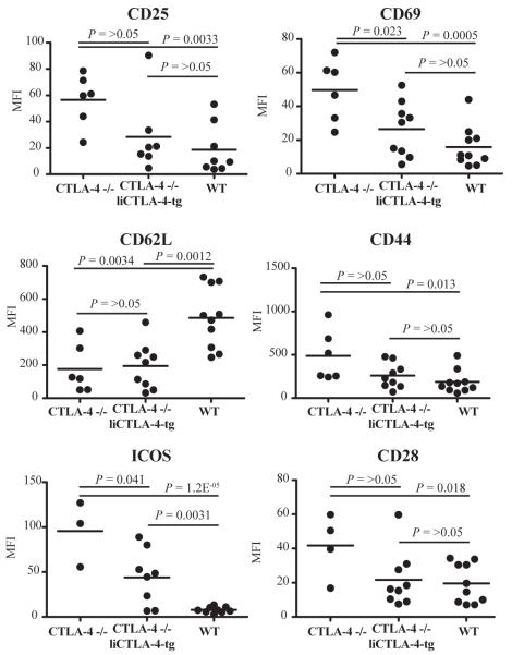
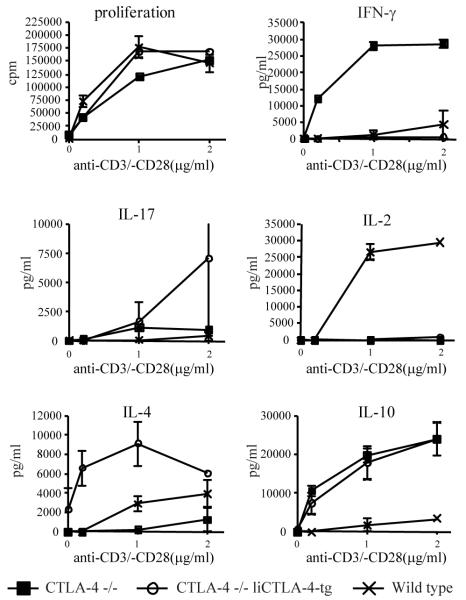
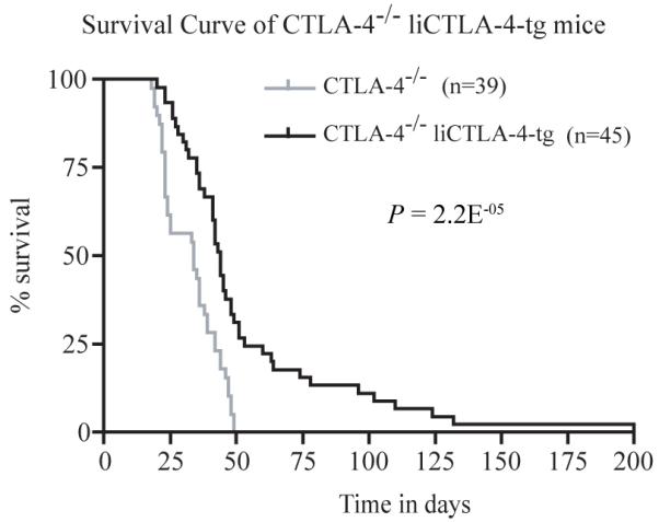
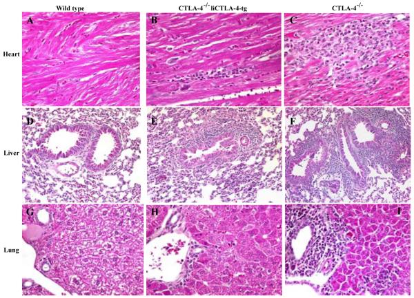
Idd5.1 regulates T1D susceptibility in nonobese diabetic (NOD) mice and has two notable candidate genes, Ctla4 and Icos. Reduced expression of one of the four CTLA-4 isoforms, ligand-independent CTLA-4 (liCTLA-4), which inhibits in vitro T cell activation and cytokine production similarly to full-length CTLA-4 (flCTLA-4), has been hypothesized to increase type 1 diabetes (T1D) susceptibility. However, further support of this hypothesis is required since the Idd5.1 haplotypes of the diabetes-susceptible NOD and the resistant B10 strains differ throughout Ctla4 and Icos. Using haplotype analysis and the generation of novel Idd5.1-congenic strains that differ at the disease-associated Ctla4 exon 2 single-nucleotide polymorphism, we demonstrate that increased expression of liCTLA-4 correlates with reduced T1D susceptibility. To directly assess the ability of liCTLA-4 to modulate T1D, we generated liCTLA-4-transgenic NOD mice and compared their diabetes susceptibility to nontransgenic littermates. NOD liCTLA-4-transgenic mice were protected from T1D to the same extent as NOD.B10 Idd5.1-congenic mice, demonstrating that increased liCTLA-4 expression alone can account for disease protection. To further investigate the in vivo function of liCTLA-4, specifically whether liCTLA-4 can functionally replace flCTLA-4 in vivo, we expressed the liCTLA-4 transgene in CTLA-4(-/-) B6 mice. CTLA-4(-/-) mice expressing liCTLA-4 accumulated fewer activated effector/memory CD4(+) T cells than CTLA-4(-/-) mice and the transgenic mice were partially rescued from the multiorgan inflammation and early lethality caused by the disruption of Ctla4. These results suggest that liCTLA-4 can partially replace some functions of flCTLA-4 in vivo and that this isoform evolved to reinforce the function of flCTLA-4.


Figures

Figure 1
Figure 1. Multiple Idd5.1 haplotypes in inbred mouse strains influence resistance and susceptibility to T1D
(A) Haplotype analysis of the Idd5.1 region was performed in DNA from eleven strains of mice. For the SNPs analyzed, those shaded in dark grey have a B10 allele and those shaded in light grey have a NOD allele. Ctla4 and Icos exonic SNPs are shown in the expanded portions of the figure where intermediate shading indicates the CAST-specific SNPs. The frequency of diabetes was monitored in (NOD.CAST Idd5.1 X NOD) F2 (B) and (NOD X NOD.SWR Idd5.1) F2 (C) cohorts as described in the Materials and Methods. As compared to littermates homozygous for the NOD haplotype at Idd5.1, homozygous (P = 0.0002) and heterozygous (P = 0.0017) NOD.CAST Idd5.1 mice are protected from diabetes while NOD.SWR Idd5.1 mice (homozygotes and heterozygotes) have a NOD-like frequency. The frequency of diabetes was compared between strains with the Kaplan-Meier log-rank test
Figure 2
Figure 2. Differential expression of liCTLA-4 mRNA and protein in Idd5.1 congenic strains
mRNA expression levels in NOD, NOD.SWR Idd5.1, NOD.CAST Idd5.1, and NOD.B10 Idd5.1 splenocytes were compared for (A) liCTLA-4, (B) flCTLA-4, (C) liCTLA-4 from multiple experiments normalized to flCTLA-4 mRNA for each individual mouse, and (D) flICOS. Spleen cells were activated in vivo for 90 min using 1 μg of anti-CD3 (clone 145-2C11) (A, B and D) or with one of two activation protocols (90 min/1 μg or 6 h/5 μg in vivo) (C) prior to mRNA isolation. The ΔCt for each sample was determined using the following formula: Cttest gene-CtB2M. Data are presented as the mean +/- SE. In C, ΔΔCt values were determined following the normalization of average Ct values using the following formula: CtflCTLA-4-CtliCTLA-4. Quantitative PCR data were evaluated using an unpaired T test. (E) Western blot analysis of liCTLA-4 and flCTLA-4 protein expression in spleen cells from NOD, NOD.SWR Idd5.1, NOD.CAST Idd5.1, NOD.B10 Idd5.1, and B6 mice. Lysates from cells were immunoblotted with anti-CTLA-4 antibody C-19 (Santa Cruz Biotech).
Figure 3
Figure 3. liCTLA-4 mRNA and protein expression in liCTLA-4 transgenic mice
(A) Schematic structure of the liCTLA-4 vector construct. liCTLA-4 was subcoloned into the EcoR1 site of pBluescript vector encoding 5′ CD2 promoter and 3′ CD2 enhancer. (B) liCTLA-4 and flCTLA-4 mRNA expression was compared between non-transgenic and liCTLA-4 transgenic mice. Spleen cells were activated in vivo for 90 minutes using 1 μg of anti-CD3 (clone 145-2C11) prior to mRNA isolation. The ΔCt for each sample of spleen cells was determined using the following formula: CtliCTLA-4-CtB2M. Data are presented as the mean ΔCt +/- SE. Quantitative PCR data were evaluated using an unpaired T test. (C) For Western analysis, 2 × 10⁶ spleen cells were immunoblotted with anti-CTLA-4 antibody (C-19, Santa Cruz Biotech).
Figure 4
Figure 4. T cell proliferation, IL-17 and IFN-γ production, and diabetes frequency are reduced in NOD liCTLA-4 transgenic mice
(A) Lymph node T cells from NOD liCTLA-4 tg and non-tg littermates were stimulated with the indicated concentrations of anti-CD3 antibody and 1 μg /ml of anti-CD28. T cell proliferation was measured with a [3H] thymidine incorporation assay and the data are presented as the mean cpm of triplicate wells. Culture supernatants were assayed by ELISA in triplicate for detection of IFN-γ and IL-17. IL-10 and IL-4 were not detected in the supernatants (data not shown). Data represent the average of three independent experiments with at least two mice in each group for each experiment. A 95% level of confidence was used to calculate the error bars. (B) NOD liCTLA-4 tg mice are protected from diabetes when compared to their non-transgenic littermate controls (P = 0.0115) The frequency of diabetes in NOD liCTLA-4 tg mice is similar to that in NOD.B10 Idd5.1 congenic mice (C), which are protected as compared to NOD mice (P = 0.0095). The frequency of diabetes was compared between strains with the Kaplan-Meier log-rank test.
Figure 5
Figure 5. CTLA-4-/- liCTLA-4 tg T cells share most cellular phenotypes with WT T cells
Average MFI of the activation markers CD25, CD69, CD62L, CD44, ICOS and CD28, in CTLA-4-/-, CTLA-4-/- liCTLA-4 tg, wild type mice (3-7 weeks of age, both sexes). Note that group sizes are not equal for each activation marker since not all antibody reagents were available each day that cells from individual mice were evaluated. P values shown in the figure were determined using the unpaired T test.
Figure 6
Figure 6. Decreased IFN-γ and IL-2 with increased IL-17, IL-4, and IL-10 production by CTLA-4-/- liCTLA-4 tg cells
Lymph node T cells from male and female 3-week-old CTLA-4-/-, CTLA-4-/- liCTLA-4 tg and WT mice were stimulated with the indicated concentrations of anti-CD3 and 1 μg /ml of anti-CD28 antibody. T cell proliferation was measured by [3H]thymidine incorporation and represented as mean CPM of triplicates (upper left). Culture supernatants were assayed by ELISA in triplicate for detection of IFN-γ, IL-4, IL-10, IL-2, and IL-17 after 48 hours. Data are representative of 3 independent experiments with 4 mice in each group. A 95% level of confidence was used to calculate the error bars.
Figure 7
Figure 7. liCTLA-4 prevents early lethality from CTLA-4 deficient mice
Survival curves for CTLA-4-/- mice (grey line) and CTLA-4-/- liCTLA-4 tg mice (black line). Survival was compared with the Kaplan-Meier log rank test.
Figure 8
Figure 8. Histopathological findings in WT, CTLA-4-/- liCTLA-4 tg and CTLA-4-/- mice reveals that CTLA-4-/- liCTLA-4-tg mice have an intermediate phenotype
Representative findings in heart (A, B and C), lung (D, E and F), and liver (G, H and I) tissues are shown. WT mice show no inflammation; CTLA-4-/- liCTLA-4-tg mice have mild myocarditis, hepatitis and increased BALT. CTLA-4-/- mice have severe myocarditis, hepatitis and large BALT. Hematoxylin and eosin, original magnifications: A-C, G-I, 160X; D-F, 80X.

References

    1. Leiter EH. Nonobese diabetic mice and the genetics of diabetes susceptibility. Curr Diab Rep. 2005;5:141–148. - PubMed
    1. Maier LM, Wicker LS. Genetic susceptibility to type 1 diabetes. Curr Opin Immunol. 2005;17:601–608. - PubMed
    1. Todd JA, Walker NM, Cooper JD, Smyth DJ, Downes K, Plagnol V, Bailey R, Nejentsev S, Field SF, Payne F, Lowe CE, Szeszko JS, Hafler JP, Zeitels L, Yang JH, Vella A, Nutland S, Stevens HE, Schuilenburg H, Coleman G, Maisuria M, Meadows W, Smink LJ, Healy B, Burren OS, Lam AA, Ovington NR, Allen J, Adlem E, Leung HT, Wallace C, Howson JM, Guja C, Ionescu-Tirgoviste C, Simmonds MJ, Heward JM, Gough SC, Dunger DB, Wicker LS, Clayton DG. Robust associations of four new chromosome regions from genome-wide analyses of type 1 diabetes. Nat Genet. 2007;39:857–864. - PMC - PubMed
    1. Vella A, Cooper JD, Lowe CE, Walker N, Nutland S, Widmer B, Jones R, Ring SM, McArdle W, Pembrey ME, Strachan DP, Dunger DB, Twells RC, Clayton DG, Todd JA. Localization of a type 1 diabetes locus in the IL2RA/CD25 region by use of tag single-nucleotide polymorphisms. Am J Hum Genet. 2005;76:773–779. - PMC - PubMed
    1. Smyth D, Cooper JD, Collins JE, Heward JM, Franklyn JA, Howson JM, Vella A, Nutland S, Rance HE, Maier L, Barratt BJ, Guja C, Ionescu-Tirgoviste C, Savage DA, Dunger DB, Widmer B, Strachan DP, Ring SM, Walker N, Clayton DG, Twells RC, Gough SC, Todd JA. Replication of an association between the lymphoid tyrosine phosphatase locus (LYP/PTPN22) with type 1 diabetes, and evidence for its role as a general autoimmunity locus. Diabetes. 2004;53:3020–3023. - PubMed

Publication types

MeSH terms