. 2009 Oct;136(20):3495-504.
doi: 10.1242/dev.036087.

Sonic hedgehog-dependent synthesis of laminin alpha1 controls basement membrane assembly in the myotome

Affiliations

Sonic hedgehog-dependent synthesis of laminin alpha1 controls basement membrane assembly in the myotome

Claire Anderson et al. Development. 2009 Oct.

Abstract

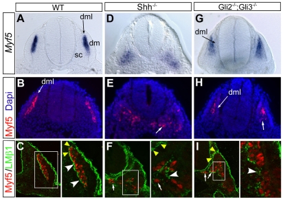
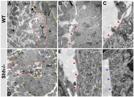
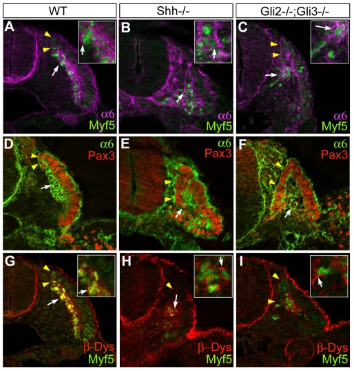
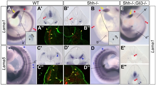
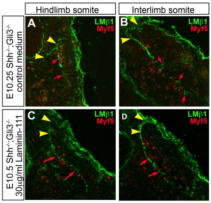
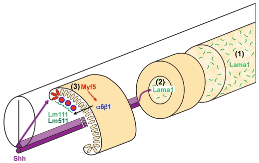
Basement membranes have essential structural and signalling roles in tissue morphogenesis during embryonic development, but the mechanisms that control their formation are still poorly understood. Laminins are key components of basement membranes and are thought to be essential for initiation of basement membrane assembly. Here, we report that muscle progenitor cells populating the myotome migrate aberrantly in the ventral somite in the absence of sonic hedgehog (Shh) signalling, and we show that this defect is due to the failure to form a myotomal basement membrane. We reveal that expression of Lama1, which encodes laminin alpha1, a subunit of laminin-111, is not activated in Shh(-/-) embryos. Recovery of Lama1 expression or addition of exogenous laminin-111 to Shh(-/-);Gli3(-/-) embryos restores the myotomal basement membrane, demonstrating that laminin-111 is necessary and sufficient to initiate assembly of the myotomal basement membrane. This study uncovers an essential role for Shh signalling in the control of laminin-111 synthesis and in the initiation of basement membrane assembly in the myotome. Furthermore, our data indicate that laminin-111 function cannot be compensated by laminin-511.


Figures

Fig. 1.
Fig. 1.
Myotome patterning defect in the absence of Shh-Gli signalling correlates with loss of a laminin-containing myotomal basement membrane. (A-I) Myf5 (red) and β1 laminin (green) expression examined by whole-mount in situ hybridisation (A,D,G) and immunohistochemistry (B,C,E,F,H,I) in rostral somites of E9.5 wild-type (A-C), Shh-/- (D-F) and Gli2-/-;Gli3-/- (G-I) mouse embryos. (A-C) Wild-type embryos. (D-F) Shh-/- embryos. (G-I) Gli2-/-;Gli3-/- embryos. Dm, dermomyotome; dml, dorso-medial lip of the dermomyotome; sc, sclerotome. White arrows show ventral Myf5-positive cells in E,H,F and I. White arrowheads indicate short fragments of assembled laminin in F,I. Yellow arrowheads indicate the basement membrane around the dorsal and ventral lips of the dermomyotome in C,F,I. Note also the ventral extension of the dml. Magnification: ×200 and ×400 (right-hand panels in C,F,I).
Fig. 2.
Fig. 2.
The myotomal basement membrane fails to form in Shh mutant embryos. (A-F) E9.5 wild-type (A-C) and Shh-/- (D-F) interlimb somites were examined by transmitted electron microscopy. (A) Wild-type somites (×1150). (B) Higher magnification (×4500) of framed area in A shows a continuous basement membrane deposited at the surface of myotomal cells (red arrowheads). (C) High magnification of framed area in B (×34,000) confirms the presence of a myotomal basement membrane (red arrowheads). (D) Shh-/- somites with myotomal cells in the ventral somite (white asterisks) (×1150). (E,F) High magnification (×34,000) of framed areas in D. (E) A basement membrane forms at the basal surface of dermomyotomal cells (red arrowheads), (F) but not at the surface of myotomal cells (blue arrowheads). The yellow dashed line indicates the dermomyotome. The red dashed line marks the myotome. dm, dermomyotome; dml, dorso-medial lip of dermomyotome; my, myotome; nt, neural tube; mt, mitochondria. Scale bars: 5 μm in D; 0.05 μm in E.
Fig. 3.
Fig. 3.
The laminin receptors, α6β1 integrin and dystroglycan, are expressed in Shh mutant embryos. (A-I) α6 integrin (purple in A-C, green in D-F) and β-dystroglycan (G-I, red) protein expression in relation to Myf5 (A-C,G-I, green) or Pax3 (D-F, red) distribution was examined by immunohistochemistry and confocal imaging in forelimb somites of E9.5 wild-type (A,D,G), Shh-/- (B,E,H), and Gli2-/-;Gli3-/- (C,F,I) embryos. (E,F) Note the increased number of Pax3-positive cells in the myotome in Shh-/- and Gli2-/-;Gli3-/-. (G-I) Note the misorientation of myotomal cells in Gli2-/-;Gli3-/- embryos (white arrow in I). Yellow arrowheads indicate receptor expression in dermomyotomal cells. White arrows show receptor expression in myotomal cells. Magnification: ×400; ×630 in insets.
Fig. 4.
Fig. 4.
Laminin α1 expression is disrupted in the absence of Shh signalling. (A-E″) Whole-mount in situ hybridisation was performed on E9.5 wild-type (A,A′,C,C′), Shh-/- (B,B′,D,D′) and Shh-/-;Gli3-/- (E,E′,E″) mouse embryos using DIG-labelled RNA probes for Lama1 (A,B,E) and Lama5 (C,D). Laminin α1 (A″) and α5 (C″) were also detected by immunofluorescence. (A,A′) Lama1 expression in wild-type embryos. (A″) Laminin α1 immunodetection in wild-type interlimb somites. (B,B′) Lama1 expression in Shh-/- embryos. Transverse sections of newly-formed (inset in A and B) and interlimb (A′,B′) somites are shown. (B″) Laminin α1 immunodetection in Shh-/- interlimb somites. (C,C′) Lama5 expression in wild-type embryos. (C″) Laminin α5 immunodetection in wild-type interlimb somites. (D,D′) Lama5 expression in Shh-/- embryos. Transverse sections of interlimb somites (C′,D′) are shown. (D″) Laminin α5 immunodetection in wild-type interlimb somites. (E,E′,E″) Lama1 expression in Gli3-/-;Shh-/- embryos. The position of transverse sections shown in E′ and E″ is indicated. Yellow arrows and red asterisks show Lama1 expression in the pre-somitic mesoderm and newly-formed somites, respectively. Red arrows indicate sites of expression affected in the absence of Shh. Blue asterisks indicate Lama5 expression in newly formed somites, and blue arrowheads show dermomyotomal expression. s, sclerotome; dm, dermomyotome. Magnification: ×250 (A-E) and ×400 (A′-E′,A″-E″).
Fig. 5.
Fig. 5.
Progressive recovery of myotomal basement membrane in Gli3-/-;Shh-/- embryos. (A-I) β1 laminin subunit (green) and Myf5 (red) protein distribution examined by immunohistochemistry and confocal imaging in somites of E10.0 Shh-/- (A-C), Shh-/-;Gli3-/- (D-F), and wild-type (G-I) embryos. (A-C) Shh-/- embryos. (D-F) Shh-/-;Gli3-/- somites. Note the recovery of epaxial Myf5 activation at all axial levels (white asterisk). (G-I) Wild-type embryos. Yellow arrowheads indicate the dermomyotomal basement membrane. Red arrows show abnormally located myotomal cells. White arrows indicate sites of partially assembled basement membrane. Magnification: ×400.
Fig. 6.
Fig. 6.
Exogenous laminin-111 restores myotomal basement membrane formation. (A-D) Laminin (green) and Myf5 (red) distribution in E10.5 Shh-/-;Gli3-/- embryos cultured for 12 hours in control medium (A,B) or in the presence of laminin-111 (30 μg/ml) (C,D). (A) No myotomal basement membrane forms in Shh-/-;Gli3-/- hindlimb somites, whereas (B) a higher degree of organisation is observed in Shh-/-;Gli3-/- interlimb somites in control medium. By contrast, Shh-/-;Gli3-/- hindlimb (C) and interlimb (D) somites form a myotomal basement membrane when cultured in the presence of laminin-111 (red arrows). Yellow arrowheads indicate the dermomyotomal basement membrane. Magnification: ×200.
Fig. 7.
Fig. 7.
Model for myotomal basement membrane formation. (1) Laminin α1 (green) is synthesised independently of Shh in the pre-somitic mesoderm. (2) Laminin α1 is synthesised under the control of Shh signalling (purple arrow) in the paraxial mesoderm at the time somites form and assembles with β1 and γ1 laminin chains into a laminin-111 network in the sclerotome/somitocoele. (3) Under Shh signalling, dorso-medial lip cells in the dermomyotome activate Myf5 (orange), which is required to upregulate the expression of the laminin receptor, α6β1 integrin (blue), in MPCs that delaminate and populate the myotome. Dorso-medial lip cells also synthesise laminin α5, which presumably begin to polymerise. However, laminin-511 cannot initiate myotomal basement membrane assembly. This requires laminin-111, which we hypothesise interacts with higher affinity to a receptor molecule present at the surface of MPCs through its LG domains. Once initiated, the myotomal basement membrane incorporates laminin-511 and other extracellular matrix components such as collagen IV, perlecan and nidogen to form a continuous, stable myotomal basement membrane.

References

    1. Andac, Z., Sasaki, T., Mann, K., Brancaccio, A., Deutzmann, R. and Timpl, R. (1999). Analysis of heparin, alpha-dystroglycan and sulfatide binding to the G domain of the laminin alpha1 chain by site-directed mutagenesis. J. Mol. Biol. 287, 253-264. - PubMed
    1. Anderson, C., Winder, S. J. and Borycki, A. G. (2007). Dystroglycan protein distribution coincides with basement membranes and muscle differentiation during mouse embryogenesis. Dev. Dyn. 236, 2627-2635. - PubMed
    1. Bader, B. L., Smyth, N., Nedbal, S., Miosge, N., Baranowsky, A., Mokkapati, S., Murshed, M. and Nischt, R. (2005). Compound genetic ablation of nidogen 1 and 2 causes basement membrane defects and perinatal lethality in mice. Mol. Cell. Biol. 25, 6846-6856. - PMC - PubMed
    1. Bajanca, F., Luz, M., Duxson, M. J. and Thorsteinsdottir, S. (2004). Integrins in the mouse myotome: developmental changes and differences between the epaxial and hypaxial lineage. Dev. Dyn. 231, 402-415. - PubMed
    1. Bajanca, F., Luz, M., Raymond, K., Martins, G. G., Sonnenberg, A., Tajbakhsh, S., Buckingham, M. and Thorsteinsdottir, S. (2006). Integrin alpha6beta1-laminin interactions regulate early myotome formation in the mouse embryo. Development 133, 1635-1644. - PubMed

Publication types

MeSH terms