. 2010 Feb;9(2):228-41.
doi: 10.1128/EC.00197-09. Epub 2009 Sep 25.

Suggestive evidence for Darwinian Selection against asparagine-linked glycans of Plasmodium falciparum and Toxoplasma gondii

Affiliations

Suggestive evidence for Darwinian Selection against asparagine-linked glycans of Plasmodium falciparum and Toxoplasma gondii

G Guy Bushkin et al. Eukaryot Cell. 2010 Feb.

Abstract

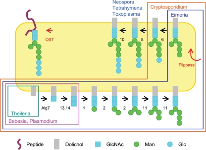
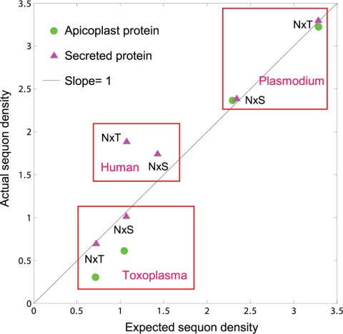
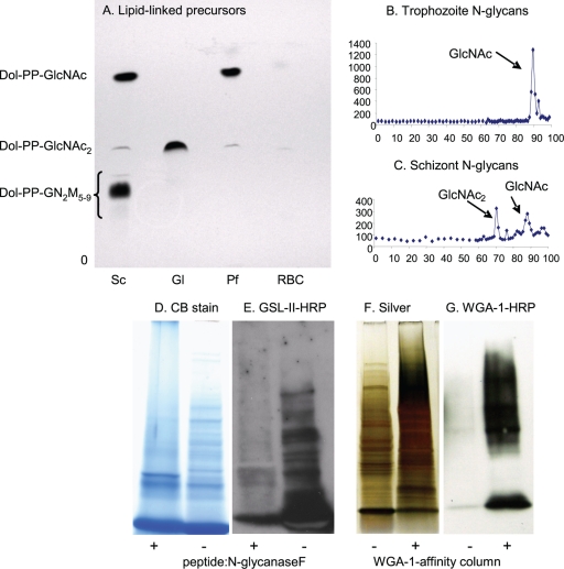
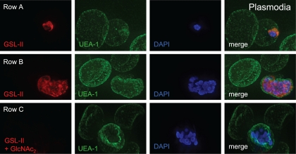
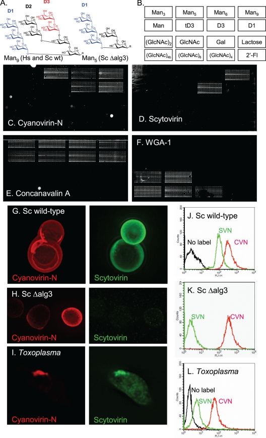
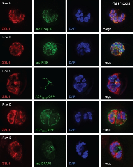
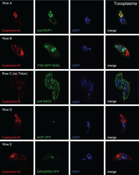
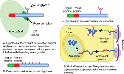
We are interested in asparagine-linked glycans (N-glycans) of Plasmodium falciparum and Toxoplasma gondii, because their N-glycan structures have been controversial and because we hypothesize that there might be selection against N-glycans in nucleus-encoded proteins that must pass through the endoplasmic reticulum (ER) prior to threading into the apicoplast. In support of our hypothesis, we observed the following. First, in protists with apicoplasts, there is extensive secondary loss of Alg enzymes that make lipid-linked precursors to N-glycans. Theileria makes no N-glycans, and Plasmodium makes a severely truncated N-glycan precursor composed of one or two GlcNAc residues. Second, secreted proteins of Toxoplasma, which uses its own 10-sugar precursor (Glc(3)Man(5)GlcNAc(2)) and the host 14-sugar precursor (Glc(3)Man(9)GlcNAc(2)) to make N-glycans, have very few sites for N glycosylation, and there is additional selection against N-glycan sites in its apicoplast-targeted proteins. Third, while the GlcNAc-binding Griffonia simplicifolia lectin II labels ER, rhoptries, and surface of plasmodia, there is no apicoplast labeling. Similarly, the antiretroviral lectin cyanovirin-N, which binds to N-glycans of Toxoplasma, labels ER and rhoptries, but there is no apicoplast labeling. We conclude that possible selection against N-glycans in protists with apicoplasts occurs by eliminating N-glycans (Theileria), reducing their length (Plasmodium), or reducing the number of N-glycan sites (Toxoplasma). In addition, occupation of N-glycan sites is markedly reduced in apicoplast proteins versus some secretory proteins in both Plasmodium and Toxoplasma.


Figures

Fig. 1.
Fig. 1.
The present diversity of N-glycan precursors among apicomplexan parasites is likely due to secondary loss of Alg enzymes. Metazoans, fungi, plants, and algae have a complete set of Alg enzymes and so make an N-glycan precursor with 14 sugars (Glc3Man9GlcNAc2) (26, 46). Toxoplasma, Neospora, and the related ciliate Tetrahymena (15) are missing Alg enzymes that add four Man residues in the ER lumen and so make an N-glycan precursor with 10 sugars (Glc3Man5GlcNAc2). Cryptosporidium is also missing Alg8 and Alg10 and so makes an N-glycan precursor with eight sugars (Glc1Man5GlcNAc2). Eimeria is also missing Alg6 and so makes an N-glycan precursor with seven sugars (Man5GlcNAc2). Plasmodium falciparum and P. vivax, as well as Babesia, are missing all the enzymes that add Man and Glc to N-glycan precursors and so add just two sugars (GlcNAc2). Theileria is missing all of the Alg enzymes, as well as the oligosaccharyltransferase (OST), and so makes no N-glycans. With the exception of Theileria, all of the apicomplexa have four OST peptides.
Fig. 2.
Fig. 2.
Negative selection against sequons (sites of N-glycans) in Toxoplasma occurs by two mechanisms. First, because Asn is encoded by AAT/C and the coding sequences of Toxoplasma are AT poor, the density of sequons in its secreted proteins (red triangles) and nucleus-encoded apicoplast proteins (green circles) is low (12). Conversely, the AT content of Plasmodium is high, and the density of sequons in its secreted proteins and apicoplast proteins is high. Humans have an intermediate AT content and therefore an intermediate sequon density in their secreted proteins. Second, there is a decreased probability that Asn, Ser, and Thr will be positioned to form sequons rather than elsewhere in nucleus-encoded apicoplast proteins of Toxoplasma. This is shown by plotting the calculated sequon density based upon amino acid composition (x axis) versus actual sequon density (y axis) for secreted proteins and apicoplast proteins of Plasmodium and Toxoplasma. In most cases, there is no selection on sequons, so points fall on the line with a slope of 1. There is, however, negative selection against sequons with Ser or Thr in apicoplast proteins of Toxoplasma, so that the actual sequon density is about one-half the calculated density. In contrast, there is positive selection for sequons with Thr in secreted proteins of humans, so that the actual sequon density is about twice the value calculated by amino acid composition (12). Because the Toxoplasma genome is AT poor and there is additional negative selection against sequons in apicoplast proteins, nearly half of the nucleus-encoded apicoplast proteins have no sequons and so cannot contain N-linked glycans (see Fig. S1 in the supplemental material). In contrast, <10% of Plasmodium secreted and apicoplast proteins have no sequons, while ∼20% of human secreted proteins have no sequons (12).
Fig. 3.
Fig. 3.
Plasmodia metabolically labeled with tritiated glucosamine (GlcN) make N-glycan precursors and N-glycans composed of a single GlcNAc and of GlcNAc2. (A) TLC shows that Plasmodium (Pf) trophozoites (early stages) make predominantly dolichol-PP-GlcNAc with a slight amount of dolichol-PP-GlcNAc2. For comparison, Giardia (Gl), which has the same Alg enzymes as Plasmodium (Fig. 1) (46), makes predominantly dolichol-PP-GlcNAc2, while Saccharomyces (Sc) makes a mixture of dolichol-PP-GlcNAc2 and high-mannose dolichol-PP-glycans. (B) N-glycans released by PNGaseF from glycoproteins of Plasmodium trophozoites are composed predominantly of a single GlcNAc. (C) N-glycans of Plasmodium schizonts (later stages) are a mixture of GlcNAc and GlcNAc2. (D to G) Additional biochemical evidence for short N-glycans of Plasmodium is shown by blots with the GlcNAc-binding lectin GSL-II after treatment of proteins with PNGaseF and by WGA-1 blots of WGA-1-affinity-purified proteins. PNGaseF-treated Plasmodium proteins, which are stained with Coomassie blue (D), show a marked decrease in binding GSL-II (E). This result shows that GSL-II is binding to N-glycans. WGA-1-affinity markedly enriches Plasmodium proteins, which are stained with silver (F), that bind to the lectin (G). Proteins identified by mass spectroscopy after a representative WGA-1 affinity experiment are shown in Table 1.
Fig. 4.
Fig. 4.
Deconvolving micrographs of Plasmodium-infected RBCs show that the GlcNAc-binding lectin GSL-II (red) (59) is specific for plasmodia, while the fucose-binding lectin UEA-1 (green) is specific for the surface of RBCs. Row A, an early trophozoite of Plasmodium has a single nucleus stained with DAPI (blue in all micrographs) and a cup-shaped secretory system stained with GSL-II after permeabilization with nonionic detergent. Row B, schizonts (later stages) of Plasmodium have multiple nuclei and much more widespread staining with GSL-II. Row C, the labeling by GSL-II of late stage plasmodia is blocked by coincubation with GlcNAc2 (diacetyl-chitobiose). GSL-II does not bind to cytosolic proteins (see Fig. S2A in the supplemental material), but GSL-II binds to the surface of plasmodia which have been released from RBCs with saponin (see Fig. S2B).
Fig. 5.
Fig. 5.
Use of antiretroviral lectins cyanovirin-N and scytovirin, which have distinct N-glycan specificities, to argue for the presence of N-glycans derived from the parasite and the host in apical secretory vesicles of Toxoplasma. (A) Man9GlcNAc2, which is the N-glycan present on glycoproteins of host cells and wild-type Saccharomyces after Glc residues are removed by glucosidases, has three arms, which are labeled D1 to D3. Man5GlcNAc2, which is the glycan made from N-glycan precursors of Toxoplasma and Saccharomyces ΔAlg3 mutants after Glc residues are removed by glucosidases, has a single D1 arm. In chemically synthesized Man9 and Man5, the two GlcNAcs are replaced by a linker that binds the sugars to the glass slide (2, 42). (B) Map of spotted sugars in panels C to F. (C) Alexafluor-labeled cyanovirin-N binds to biosynthetic Man5, Man9, and both D1 and D3 arms of Man9, which have been applied to glass slides. (D) In contrast, scytovirin only binds to Man9 and its D3 arm but does not bind to Man5. (E) The plant lectin concanavalin A binds to the entire set of mannose sugars, which are arrayed on a glass slide. (F) WGA-1, which is specific for GlcNAc, fails to bind to mannose sugars. (G) Wild-type Saccharomyces, which makes N-glycans based upon a Man9GlcNac2 precursor, is labeled well by both cyanovirin-N (red) and scytovirin (green). (H) In contrast, the Saccharomyces ΔAlg3 mutant, which makes N-glycans based upon a Man5GlcNac2 precursor, is labeled with cyanovirin-N (red) but not with scytovirin (green). (I) Cyanovirin-N and scytovirin both label the apical end of a Toxoplasma tachyzoite which has been released from a monolayer of human foreskin fibroblasts. (J to L) The binding of cyanovirin-N and scytovirin to wild-type Saccharomyces (J), the Saccharomyces ΔAlg3 mutant (K), and Toxoplasma (L) were measured using flow cytometry. These results suggest that Toxoplasma N-glycans are composed of a mix of Man5GlcNAc2 and Man9GlcNAc2, as recently proposed (19).
Fig. 6.
Fig. 6.
Deconvolving micrographs of Plasmodium-infected RBCs show that GSL-II, which binds to Plasmodium N-glycans, colocalizes with rhoptries (row A) and the ER (row B) but does not colocalize with apicoplasts (row C), the parasitophorous vacuole (row D), or the food vacuole (row E). In each case GSL-II is labeled red with Alexafluor 594, while antibodies labeled green with Alexafluor 488 are against RhopH3 (rhoptries), Pf39 (ER), and food vacuole (DPAP1) (14, 30, 51). Alternatively, GFP is targeted to apicoplasts (ACPleader-GFP) and the parasitophorous vacuole (ACPsignal-GFP) (55). Control experiments with anti-GFP antibodies (see Fig. S4 in the supplemental material) show that both apicoplasts and parasitophorous vacuoles are accessible to exogenous probes.
Fig. 7.
Fig. 7.
Deconvolving micrographs of Toxoplasma tachyzoites show that cyanovirin-N, which binds to Toxoplasma N-glycans, colocalizes with rhoptries (row A), the ER (row B), and the plasma membrane (row C) but does not colocalize with apicoplasts (row D) and the Golgi apparatus (row E). In each case cyanovirin-N is labeled red with Alexafluor 594, while antibodies labeled green with Alexafluor 488 are against ROP1 (rhoptries) and SAG1 (plasma membrane). Alternatively, fluorescent proteins are targeted to apicoplasts (ACP-YFP), the ER (P30-GFP-HDEL), and the Golgi apparatus (GRASP55-YFP). Control experiments with anti-GFP antibodies (see Fig. S5 in the supplemental material) show that both apicoplasts and the Golgi apparatus are accessible to exogenous probes.
Fig. 8.
Fig. 8.
Model summarizing our hypothesis and results with Plasmodium and Toxoplasma. (A) We hypothesized that bulky N-glycans might interfere with threading of nucleus-encoded apicoplast proteins into the organelle. (B) We found that Plasmodium markedly reduces the length of its N-glycans without reducing the density of sites of N-linked glycosylation. (C) In contrast, Toxoplasma has longer N-glycans but dramatically reduces the density of sites of N-glycans (sequons), so that nearly half of the Toxoplasma nucleus-encoded apicoplast proteins have no sequons and so contain no N-glycans. (D) While secreted proteins are N glycosylated, sites of N-glycans (sequons) on nucleus-encoded apicoplast proteins of Plasmodium and Toxoplasma remain unoccupied.

References

    1. Abrahamsen M. S., Templeton T. J., Enomoto S., Abrahante J. E., Zhu G., et al. 2004. Complete genome sequence of the apicomplexan, Cryptosporidium parvum. Science 304:441–445 - PubMed
    1. Adams E. W., Ratner D. M., Bokesch H. R., McMahon J. B., O'Keefe B. R., Seeberger P. H. 2004. Oligosaccharide and glycoprotein microarrays as tools in HIV glycobiology; glycan-dependent gp120/protein interactions. Chem. Biol. 11:875–881 - PubMed
    1. Altschul S. F., Madden T. L., Schaffer A. A., Zhang J., Zhang Z., Miller W., Lipman D. J. 1997. Gapped BLAST and PSI-BLAST: a new generation of protein database search programs. Nucleic Acids Res. 25:3389–3402 - PMC - PubMed
    1. Aurrecoechea C., Brestelli J., Brunk B. P., Dommer J., Fischer S., et al. 2009. PlasmoDB: a functional genomic database for malaria parasites. Nucleic Acids Res. 37:D539–D543 - PMC - PubMed
    1. Banerjee S., Vishwanath P., Cui J., Kelleher D. J., Gilmore R., Robbins P. W., Samuelson J. 2007. Evolution of quality control of protein-folding in the ER lumen. Proc. Natl. Acad. Sci. U. S. A. 104:11676–11681 - PMC - PubMed

Publication types

MeSH terms

LinkOut - more resources