Review
. 2009 Oct;88(10):877-93.
doi: 10.1177/0022034509343280.

The impact of fluoride on ameloblasts and the mechanisms of enamel fluorosis

Affiliations
Review

The impact of fluoride on ameloblasts and the mechanisms of enamel fluorosis

A L J J Bronckers et al. J Dent Res. 2009 Oct.

Abstract

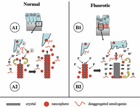
Intake of excess amounts of fluoride during tooth development cause enamel fluorosis, a developmental disturbance that makes enamel more porous. In mild fluorosis, there are white opaque striations across the enamel surface, whereas in more severe cases, the porous regions increase in size, with enamel pitting, and secondary discoloration of the enamel surface. The effects of fluoride on enamel formation suggest that fluoride affects the enamel-forming cells, the ameloblasts. Studies investigating the effects of fluoride on ameloblasts and the mechanisms of fluorosis are based on in vitro cultures as well as animal models. The use of these model systems requires a biologically relevant fluoride dose, and must be carefully interpreted in relation to human tooth formation. Based on these studies, we propose that fluoride can directly affect the ameloblasts, particularly at high fluoride levels, while at lower fluoride levels, the ameloblasts may respond to local effects of fluoride on the mineralizing matrix. A new working model is presented, focused on the assumption that fluoride increases the rate of mineral formation, resulting in a greater release of protons into the forming enamel matrix.


Figures

Figure 1.
Figure 1.
Cyst formation after single injection of a high dose of fluoride. (A) Hamster first maxillary molar tooth germ, at post-natal day 4, from an animal injected with 9 mg F/kg body weight and killed 24 hrs later. Undecalcified, hematoxylin-eosin staining (25x). Cysts have formed near the cervical area (ceC) at early-secretory stages and coronally (coC) at late-secretory/transitional stages. Enamel surface under the cysts is intensely mineralized. Fully secretory ameloblasts (fsa) are not overtly affected, but a thin weak response line (position indicated by arrowheads) runs almost parallel to the surface through secretory-stage enamel (E). At the right cusp, this line connects the bases of both coronal and cervical cysts. Late-secretory-stage cells in the left cusp are not affected (Lyaruu et al., 2006). (B) Higher magnification of boxed area with double-response in adjacent section stained with toluidine blue (400x). Between arrows is the pre-exposure (‘pre’) secretory enamel that hypermineralized during exposure and stained less intensely with toluidine blue. The enamel layer secreted after the F peak insult (‘post’) by secretory ameloblasts (SA) is less mineralized and stains darker with toluidine blue. Fig. 4d illustrates a similar area with a sharp transition of hyper- to hypomineralized enamel at the ultrastructural level. Psa, pre-secretory ameloblasts; fsa, fully secretory ameloblasts; ta, transitional ameloblasts; ma, maturation ameloblasts; d, dentin; o, odontoblasts, p, pulp.
Figure 2.
Figure 2.
Normal amelogenesis (A) and amelogenesis 24 hrs after an acute high exposure to fluoride (B). (A) Schematic drawing of normal amelogenesis in an imaginary cusp of a molar. D, dentin; E, enamel. Increasing greyness in enamel represents increasing mineral content. Successive stages of development from bottom to top. Aprismatic enamel with small crystallites is produced by early- and late-secretory ameloblasts. The bulk of (inner) enamel consists of prisms containing large crystals and is deposited by fully differentiated ameloblasts. (B) Amelogenesis 24 hrs after injection of 9 mg F/kg. Fluoride induces an (inner) hypermineralized layer (black line, white arrows) in fully secretory-stage enamel, running almost parallel to the surface of the enamel. It represents the mineralization front 24 hrs earlier at the time of fluoride injection. A later-formed (outer) layer is a hypomineralized layer (white line, black arrows); together, these lines form the double-response typical of fluoride. Two areas of intense hypermineralization are formed at both ends of these lines: one where the lines intersect the enamel-dentin junction in the inner aprismatic enamel, the other where the lines intersect the enamel surface with outer aprismatic enamel below late-secretory and transitional ameloblasts. Cyst formation occurs only under some groups of transitional ameloblasts (coronal cyst, coC) and early-secretory ameloblasts (cervical cyst, ceC) that detach from enamel surface. Maturation ameloblasts seem structurally unaltered. Fully secretory-stage ameloblasts recover completely after 24 hrs; only the double-response lines are reminiscent of the insult.
Figure 3.
Figure 3.
Permanent lesions induced by chronic or acute exposure to fluoride found or expected in erupted fluorotic enamel. During amelogenesis, the tooth was chronically exposed to low-dose fluoride in drinking water (A) or to a single high parenteral dose of fluoride (B). Black represents the fully mineralized enamel, grey, hypomineralized. E, enamel; D, dentin. (A) Characteristic of low chronic doses of fluoride acting on the maturation stage is development of the hypomineralized subsurface area (grey), and a thin hypermineralized outer surface layer (black). Multiple weak hypo- and hypermineralized (grey) lines run through the enamel, the double-response lines, formed at the secretory stage. The enamel-dentin junction also contains a double-response (not shown). (B) Post-eruptive defects after a single high plasma peak level of fluoride (9 mg F/kg injection) are deep (hypothetical) and shallow pits that result from sub-ameloblastic cysts formed by damaged early- and late-secretory ameloblasts. The altered mineralization pattern seen in the double-response line partially recovers during post-exposure maturation, but remains hypomineralized. Maturation stages may also be affected (indicated by question mark) by chronic low but sustained levels of fluoride over time released by bone remodeling after clearance of F plasma peak levels.
Figure 4.
Figure 4.
Ultrastructural micrographs of hamster molar tooth germs cultured for 24 hrs in the presence of 5 mg F/L (A,B) followed by a 24-hour recovery in fluoride-free media (C,D). (A) Fluorotic matrix (FM) fails to mineralize, whereas the initial aprismatic enamel (FE) deposited prior to fluoride exposure hypermineralizes intensely (about 15-fold of the pre-exposure control values). All the enamel crystals are laterally fused [see Lyaruu et al. (1989a) for details]. D, dentin. Original magnification: 26,400x. (B) Prismatic secretory enamel (FE) situated more occlusally also hypermineralizes, but to a lesser extent (about five-fold of control levels). Although hypermineralized, there are no lateral enamel crystal fusions. The new fluorotic matrix (FM) fails to mineralize. Original magnification: 34,000x. (C) Same enamel region as that shown in Fig. 4A after recovery. The fluorotic matrix has recovered (RE) after 24-hour culture in fluoride-free media. The new crystals are thin compared with the fluorotic crystals. The crystals show preferred orientation within the matrix, but are not continuous with crystals in the fluorotic enamel (FE). This is a potential fracture plane after tooth eruption. D, Dentin. Original magnification: 20,500x. (D) Enamel region comparable with that shown in 4B, but after a 24-hour “recovery” culture in fluoride-free medium. The fluorotic matrix has recovered (RE), i.e., mineralized. The new crystals are thin and ribbon-like (upper inset, side view of one crystal between arrowheads) and initiate and extend from the fluorotic enamel (FE) crystals. The degree of hypermineralization of the fluorotic enamel crystals did not change during the recovery period (lower inset, side view of one crystal between arrowheads; same magnification as upper inset; note difference in thickness with upper inset). D = dentin. Original magnification: 20,500x. Insets: 100,000x.
Figure 5.
Figure 5.
Proposed molecular mechanism for enamel fluorosis. (A) Early-secretory amelogenesis in non-fluorotic conditions (adapted from Fincham et al., 1995). (A1) Light-microscope level. Boxed area is enlarged in A2 and represents crystal formation at the molecular level. (A2) Tomes’ process (1) secretes a secretory vesicle containing amelogenins. At neutral pH, amelogenins (for simplicity, drawn as a thread) form nanospheres (2) that adhere to the surfaces of growing crystals and foster preferential crystal growth in length, but reduce growth in width. Crystal growth generates protons that need to be neutralized to drive further crystal growth. Amelogenins bind and neutralize protons (3). The result of this action is an increase in crystal length, seen at the right side of this picture. (B) Disruption of secretory amelogenesis by fluoride as seen in vitro. (B1) Light-microscopic level. The boxed area in B1 is magnified in B2. (B2) Fluoride primarily accelerates crystal growth in thickness (left drawing). This forms the hypermineralized line, the initial event of the double-response. Accelerated mineral deposition strongly enhances proton production (right side drawing) that cannot be buffered by the available amelogenins. Amelogenin nanospheres at acid pH deaggregate and detach from the crystal surface. Also, newly secreted matrix will not form nanospheres at low pH and remains monomeric (fluid). Control of preferential crystal growth in length is then lost at acid pH, and newly secreted matrix will not foster crystal growth until neutral pH is restored. This layer represents the hypomineralized line, the outer component of the double-response event. These fluoride-related changes in the matrix could further alter ameloblast cell function.

References

    1. Andersen L, Richards A, Care AD, Andersen HM, Kragstrup J, Fejerskov O. (1986). Parathyroid glands, calcium, and vitamin D in experimental fluorosis in pigs. Calcif Tissue Int 38:222-226 - PubMed
    1. Angmar-Månsson B, Whitford GM. (1982). Plasma fluoride levels and enamel fluorosis in the rat. Caries Res 16:334-339 - PubMed
    1. Angmar-Månsson B, Whitford GM. (1984). Enamel fluorosis related to plasma F levels in the rat. Caries Res 18:25-32 - PubMed
    1. Angmar-Månsson B, Whitford GM. (1985). Single fluoride doses and enamel fluorosis in the rat. Caries Res 19:145-152 - PubMed
    1. Angmar-Månsson B, Ericsson Y, Ekberg O. (1976). Plasma fluoride and enamel fluorosis. Calcif Tissue Res 22:77-84 - PubMed

Publication types

LinkOut - more resources