. 2009 Nov;37(21):7290-301.
doi: 10.1093/nar/gkp793.

The HSV-1 ICP27 RGG box specifically binds flexible, GC-rich sequences but not G-quartet structures

Affiliations

The HSV-1 ICP27 RGG box specifically binds flexible, GC-rich sequences but not G-quartet structures

Kara A Corbin-Lickfett et al. Nucleic Acids Res. 2009 Nov.

Abstract

Herpes simplex virus 1 (HSV-1) protein ICP27, an important regulator for viral gene expression, directly recognizes and exports viral RNA through an N-terminal RGG box RNA binding motif, which is necessary and sufficient for RNA binding. An ICP27 N-terminal peptide, including the RGG box RNA binding motif, was expressed and its binding specificity was analyzed using EMSA and SELEX. DNA oligonucleotides corresponding to HSV-1 glycoprotein C (gC) mRNA, identified in a yeast three-hybrid analysis, were screened for binding to the ICP27 N-terminal peptide in EMSA experiments. The ICP27 N-terminus was able to bind most gC substrates. Notably, the ICP27 RGG box was unable to bind G-quartet structures recognized by the RGG domains of other proteins. SELEX analysis identified GC-rich RNA sequences as a common feature of recognition. NMR analysis of SELEX and gC sequences revealed that sequences able to bind to ICP27 did not form secondary structures and conversely, sequences that were not able to bind to ICP27 gave spectra consistent with base-pairing. Therefore, the ICP27 RGG box is unique in its recognition of nucleic acid sequences compared to other RGG box proteins; it prefers flexible, GC-rich substrates that do not form stable secondary structures.


Figures

Figure 1.
Figure 1.
Schematic of the ICP27 functional domains and bacterial expression of the ICP27 N-terminal 160 amino acids. (A) ICP27 functional domains include an N-terminal leucine-rich region (LRR), acidic region (D/E), a nuclear localization signal (NLS), arginine-/glycine-rich RGG box (RGG) and a second arginine-rich region (R2.) The C-terminus contains three predicted hnRNP K homology (KH) domains and a zinc finger-like motif (CCHC). The region of the ICP27 N-terminus expressed in E. coli is underlined. (B) Codon optimized ICP27 N-terminal peptide was expressed in Rosetta E. coli with a C-terminal His tag. Expressed protein was purified using a Nickel column under native conditions. Elution fractions were collected from the Nickel column and fractions 1 through 8 were run on a 10–20% SDS–PAGE gradient gel and stained with Coomassie Blue. (C) Glycoprotein C (gC) gene sequence identified in the yeast three hybrid screen with HSV-1 UL 43 and gC RNA and full-length ICP27. DNA sequence of the 294-nt region of the gC gene identified in a yeast three-hybrid screen interacting with ICP27. This DNA sequence corresponds to nucleotides 96 946–97 239 of the KOS HSV-1 gC mRNA sequence that were identified interacting with ICP27. Sequences underlined and numbered denote the overlapping 30-mer gC DNA oligonucleotides used in EMSA with the ICP27 N-terminal peptide.
Figure 2.
Figure 2.
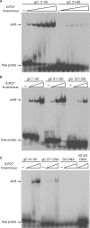
The ICP27 N-terminus binds to a majority of gC DNA sequences but not a G-quartet DNA sequence. (A) A 20 fmol of radiolabeled gC DNA oligonucleotides gC 11–40 and gC 31–60 (see figure for sequence) were either incubated with no protein or with 0.5 to 20 µM of the ICP27 N-terminal peptide. Samples were electrophoresed on a prerun acrylamide gel and dried gels were exposed to film. Arrows indicate the migration of free probe and the shift due to ICP27 N-terminal peptide binding. (B) A 20 fmol of radiolabeled gC DNA oligonucleotides gC 1–30, gC 91–120 or gC 121–150 were incubated with no protein or 0.5 to 12.5 µM of the ICP27 N-terminal peptide. Samples were electrophoresed and dried gels were exposed to film. (C) A 20 fmol of radiolabeled gC DNA oligonucleotides gC 61–90, gC 271–294 and G4 DNA [TAGGGGTT]4 were incubated with 0.5 to 12.5 µM of the ICP27 N-terminal peptide. In the lanes denoted 4X G4 DNA (far right lanes), 80 fmol of G4 DNA were used.
Figure 3.
Figure 3.
The ICP27 N-terminus binds to gC RNA sequences and the interaction between ICP27 N-terminus and individual gC DNA sequences can be competed by other high affinity binding gC DNA sequences but not by weak binding DNA sequences. (A) A 20 fmol of radiolabeled DNA or RNA oligonucleotides corresponding to the gC 1–30 or gC 31–60 sequence (see Figure 1C for sequence) were incubated with no protein (−) or 2.5, 7.5, 22.5 and 67.5 µM of the ICP27 N-terminal peptide. Samples were electrophorsesed and dried gels were exposed to film. Arrows indicate the position of free and shifted probes. (B) A 20 fmol of radiolabeled gC oligonucleotide gC 71–100 was incubated with no protein (lane 1), 5 µM of the ICP27 N-terminal peptide (lane 2) or 5 µM of the ICP27 N-terminal peptide with 5× (100 fmol), 10× (200 fmol) or 50× (1000 fmol) of nonradioactive competitor gC DNA oligonucleotides (gC 31–60, gC 271–294 or gC 11–40.) Samples were electrophoresed and dried gels were exposed to film. Arrows indicate the position of the free and shifted probes.
Figure 4.
Figure 4.
mfold analysis and representative predicted secondary structures of selected gC DNA sequences. All gC 30-mer DNA oligonucleotides were analyzed by mfold using the DNA mfold server version 3.2 (35). Default constraints were used which included a folding temperature of 37°C and an upper bound of 10 on the number of computed foldings. (A through H) The predicted mfold structure with the lowest ΔG in kcal/mol for eight out of the nineteen gC DNA sequences (Table 1) (A) gC 1–30, (B) gC 11–40, (C) gC 91–120, (D) gC 151–180, (E) gC 31–60, (F) gC 191–210, (G) gC 121–150 and (H) gC 211–240.
Figure 5.
Figure 5.
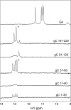
1D 1H spectra of G-quartet DNA and selected gC 30-mer DNA oligonucleotides. A portion of the 1D 1H spectra (9–14 p.p.m.) collected on 0.5 mM G4 DNA [TTGGGGTT]4, gC 191–220, gC 91–120, gC 31–60, gC 11–40 and gC 1–30 at 25°C in 50 mM Tris pH 8, 150 mM KCl and 10% D2O. Spectra were scaled using a resolved H1’ ribose signal. Asterisks (*) indicate prominent peaks detected in gC 191–220 and gC 31–60 sequences, two sequences that were not shifted by the ICP27 in EMSA experiments (Figure 2).
Figure 6.
Figure 6.
EMSA of selected SELEX DNA oligonucleotides. A 20 fmol of radiolabeled SELEX 20-mer DNA oligonucleotides (see Table 2) or the gC 11–40 DNA oligonucleotide were either incubated no protein or with 2.5–62.5 µM of the ICP27 N-terminal peptide. A 62.5 µM of the ICP27 N-terminal peptide was used in the (+) gC 11–40 lanes. Samples were electrophoresed on a prerun acrylamide gel with Tris Acetate Buffer and dried gels were exposed to film. Arrows indicate the migration of free probe and the shift due to ICP27 N-terminal peptide binding.
Figure 7.
Figure 7.
mfold analysis and representative predicted secondary structures of selected SELEX RNA sequences. All SELEX RNA sequences were submitted for mfold analysis using the RNA mfold version 3.2 (35,39). Default constraint settings were used including a folding temperature of 37°C. (A through G) The predicted mfold structure with the lowest ΔG in kcal/mol for seven out of the nineteen SELEX sequences identified. (A) SELEX 1, (B) SELEX 2, (C) SELEX 3, (D) SELEX 4, (E) SELEX 6, (F) SELEX 5 and (G) SELEX 13.
Figure 8.
Figure 8.
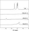
1D 1H spectra of G-quartet DNA and selected SELEX DNA sequences. A portion of the 1D 1H spectra (9–14 p.p.m.) collected on 0.5 mM G4 DNA [dTTGGGGTT]4, SELEX 13, SELEX 4 and SELEX 2 at 25°C in 50 mM Tris pH 8, 150 mM KCl and 10% D2O. Spectra were scaled using a resolved H1’ ribose signal. The asterisk (*) indicates broad peaks specifically seen in SELEX 2, a sequence that was not shifted by the ICP27 in EMSA experiments (Figure 6).

Similar articles

Cited by

References

    1. Hardy WR, Sandri-Goldin RM. Herpes simplex virus inhibits host cell splicing, and regulatory protein ICP27 is required for this effect. J. Virol. 1994;68:7790–7799. - PMC - PubMed
    1. Dai-Ju JQ, Li L, Johnson LA, Sandri-Goldin RM. ICP27 interacts with the C-terminal domain of RNA polymerase II and facilitates its recruitment to herpes simplex virus 1 transcription sites, where it undergoes proteasomal degradation during infection. J. Virol. 2006;80:3567–3581. - PMC - PubMed
    1. Sandri-Goldin RM. ICP27 mediates HSV RNA export by shuttling through a leucine-rich nuclear export signal and binding viral intronless RNAs through an RGG motif. Genes Dev. 1998;12:868–879. - PMC - PubMed
    1. Ingram A, Phelan A, Dunlop J, Clements JB. Immediate early protein IE63 of herpes simplex virus type 1 binds RNA directly. J. Gen. Virol. 1996;77((Pt 8)):1847–1851. - PubMed
    1. Chen IH, Li L, Silva L, Sandri-Goldin RM. ICP27 recruits Aly/REF but not TAP/NXF1 to herpes simplex virus type 1 transcription sites although TAP/NXF1 is required for ICP27 export. J. Virol. 2005;79:3949–3961. - PMC - PubMed

Publication types

MeSH terms