

Crystal structure of the EndoG/EndoGI complex: mechanism of EndoG inhibition
- PMID: 19783821
- PMCID: PMC2790893
- DOI: 10.1093/nar/gkp770
Crystal structure of the EndoG/EndoGI complex: mechanism of EndoG inhibition
Abstract
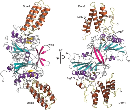
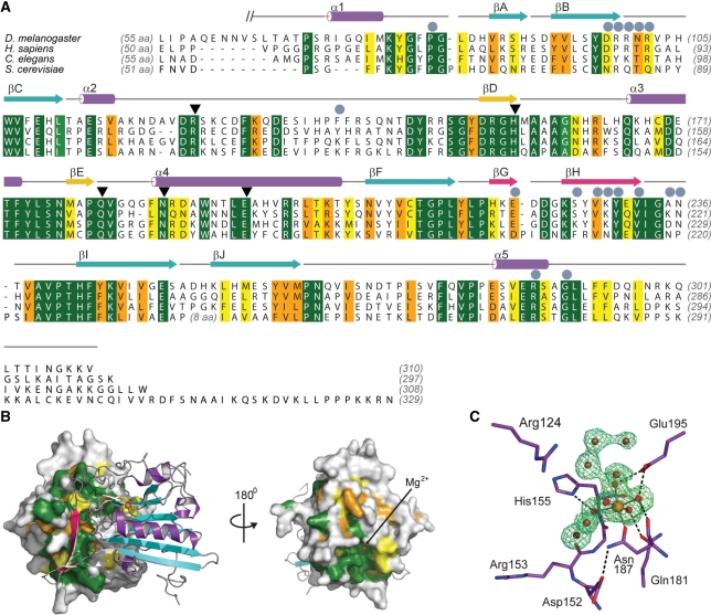
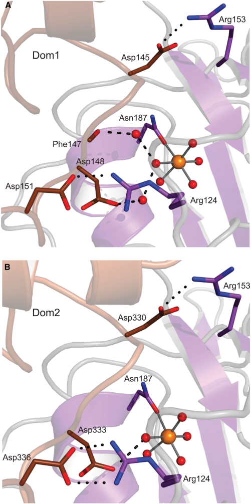
EndoG is a ubiquitous nuclease that is translocated into the nucleus during apoptosis to participate in DNA degradation. The enzyme cleaves double- and single-stranded DNA and RNA. Related nucleases are found in eukaryotes and prokaryotes, which have evolved sophisticated mechanisms for genome protection against self-antagonizing nuclease activity. Common mechanisms of inhibition are secretion, sequestration into a separate cellular compartment or by binding to protein inhibitors. Although EndoG is silenced by compartmentalization into the mitochondrial intermembrane space, a nucleus-localized protein inhibitor protects cellular polynucleotides from degradation by stray EndoG under non-apoptotic conditions in Drosophila. Here, we report the first three-dimensional structure of EndoG in complex with its inhibitor EndoGI. Although the mechanism of inhibition is reminiscent of bacterial protein inhibitors, EndoGI has evolved independently from a generic protein-protein interaction module. EndoGI is a two-domain protein that binds the active sites of two monomers of EndoG, with EndoG being sandwiched between EndoGI. Since the amino acid sequences of eukaryotic EndoG homologues are highly conserved, this model is valid for eukaryotic dimeric EndoG in general. The structure indicates that the two active sites of EndoG occupy the most remote spatial position possible at the molecular surface and a concerted substrate processing is unlikely.
Figures
Similar articles
-
The Drosophila melanogaster Gene cg4930 Encodes a High Affinity Inhibitor for Endonuclease G.J Biol Chem. 2009 Mar 27;284(13):8337-48. doi: 10.1074/jbc.M808319200. Epub 2009 Jan 7. J Biol Chem. 2009. PMID: 19129189 Free PMC article.
-
Crystal structure of the mouse endonuclease G.Biochem Biophys Res Commun. 2020 May 21;526(1):35-40. doi: 10.1016/j.bbrc.2020.03.060. Epub 2020 Mar 16. Biochem Biophys Res Commun. 2020. PMID: 32192768
-
Structural and functional characterization of mitochondrial EndoG, a sugar non-specific nuclease which plays an important role during apoptosis.J Mol Biol. 2004 Apr 23;338(2):217-28. doi: 10.1016/j.jmb.2004.02.069. J Mol Biol. 2004. PMID: 15066427
-
Crystal structure of endonuclease G in complex with DNA reveals how it nonspecifically degrades DNA as a homodimer.Nucleic Acids Res. 2016 Dec 1;44(21):10480-10490. doi: 10.1093/nar/gkw931. Epub 2016 Oct 13. Nucleic Acids Res. 2016. PMID: 27738134 Free PMC article.
-
EXOG, a novel paralog of Endonuclease G in higher eukaryotes.Nucleic Acids Res. 2008 Mar;36(4):1369-79. doi: 10.1093/nar/gkm1169. Epub 2008 Jan 10. Nucleic Acids Res. 2008. PMID: 18187503 Free PMC article.
Cited by
-
Nucleases as a barrier to gene silencing in the cotton boll weevil, Anthonomus grandis.PLoS One. 2017 Dec 20;12(12):e0189600. doi: 10.1371/journal.pone.0189600. eCollection 2017. PLoS One. 2017. PMID: 29261729 Free PMC article.
-
Dnases in health and disease.Dev Biol. 2017 Sep 1;429(1):1-11. doi: 10.1016/j.ydbio.2017.06.028. Epub 2017 Jun 28. Dev Biol. 2017. PMID: 28666955 Free PMC article. Review.
-
Anti-ROR1 scFv-EndoG as a Novel Anti-Cancer Therapeutic Drug.Asian Pac J Cancer Prev. 2018 Jan 27;19(1):97-102. doi: 10.22034/APJCP.2018.19.1.97. Asian Pac J Cancer Prev. 2018. PMID: 29373898 Free PMC article.
-
Characterization of a cell death-inducing endonuclease-like venom protein from the parasitoid wasp Pteromalus puparum (Hymenoptera: Pteromalidae).Pest Manag Sci. 2021 Jan;77(1):224-233. doi: 10.1002/ps.6011. Epub 2020 Aug 6. Pest Manag Sci. 2021. PMID: 32673424 Free PMC article.
-
Immunology and Genetic of Leishmania infantum: The Role of Endonuclease G in the Apoptosis.J Res Med Sci. 2018 Apr 26;23:36. doi: 10.4103/jrms.JRMS_705_17. eCollection 2018. J Res Med Sci. 2018. PMID: 29887904 Free PMC article. Review.
References
-
- Parrish J, Li L, Klotz K, Ledwich D, Wang X, Xue D. Mitochondrial endonuclease G is important for apoptosis in C. elegans. Nature. 2001;412:90–94. - PubMed
-
- Schafer P, Scholz SR, Gimadutdinow O, Cymerman IA, Bujnicki JM, Ruiz-Carrillo A, Pingoud A, Meiss G. Structural and functional characterization of mitochondrial EndoG, a sugar non-specific nuclease which plays an important role during apoptosis. J. Mol. Biol. 2004;338:217–228. - PubMed
-
- Buttner S, Eisenberg T, Carmona-Gutierrez D, Ruli D, Knauer H, Ruckenstuhl C, Sigrist C, Wissing S, Kollroser M, Frohlich KU, et al. Endonuclease G regulates budding yeast life and death. Mol. Cell. 2007;25:233–246. - PubMed
-
- Muro-Pastor AM, Flores E, Herrero A, Wolk CP. Identification, genetic analysis and characterization of a sugar-non-specific nuclease from the cyanobacterium Anabaena sp. PCC 7120. Mol. Microbiol. 1992;6:3021–3030. - PubMed
-
- Ball TK, Saurugger PN, Benedik MJ. The extracellular nuclease gene of Serratia marcescens and its secretion from Escherichia coli. Gene. 1987;57:183–192. - PubMed
Publication types
MeSH terms
Substances
Associated data
- Actions
LinkOut - more resources
Full Text Sources
Other Literature Sources
Molecular Biology Databases
