Comparative Study
. 2009 Nov;8(22):2136-43.
doi: 10.4161/cbt.8.22.9743. Epub 2009 Nov 7.

Inhibition of KSHV-infected primary effusion lymphomas in NOD/SCID mice by gamma-secretase inhibitor

Affiliations
Comparative Study

Inhibition of KSHV-infected primary effusion lymphomas in NOD/SCID mice by gamma-secretase inhibitor

Ke Lan et al. Cancer Biol Ther. 2009 Nov.

Erratum in

  • Correction.
    [No authors listed] [No authors listed] Cancer Biol Ther. 2021 Jun 3;22(5-6):414. doi: 10.1080/15384047.2021.1926127. Epub 2021 May 26. Cancer Biol Ther. 2021. PMID: 34039251 Free PMC article. No abstract available.

Abstract

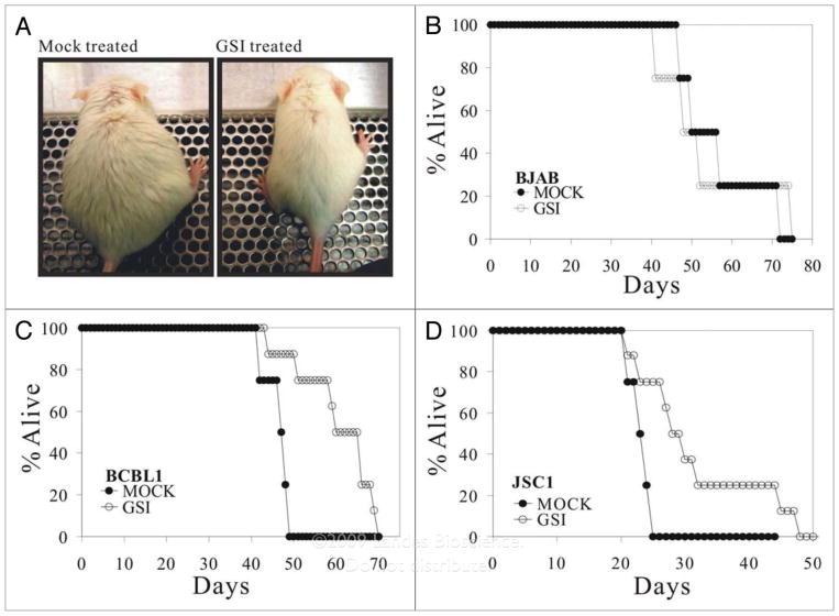
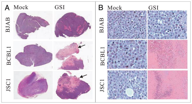
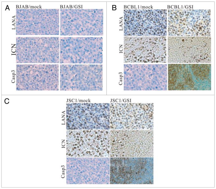
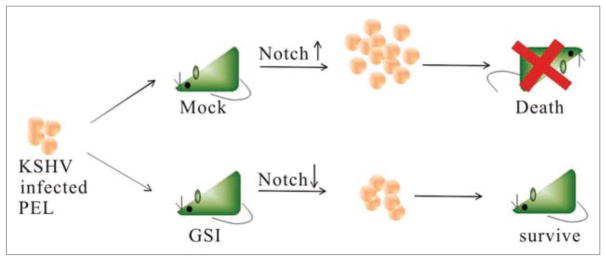
Primary effusion lymphoma (PEL) is a common cancer in AIDS patients closely associated with Kaposi's sarcoma-associated herpesvirus (KSHV). Previously, we showed that KSHV latency associated nuclear antigen (LANA) stabilizes intracellular activated Notch1 (ICN) involved in maintenance of the malignant phenotype of KSHV infected PEL cells in vitro. The gamma-secretase inhibitor (GSI) which specifically blocks the production of ICN slows down the proliferation of the KSHV infected PEL cell lines BCBL1, BC3 as well as JSC1 in vitro. In this study, we extended these studies to explore the possibility that manipulation of the Notch signaling by GSI would prevent the growth of the PEL tumors in vivo. We observed that the onset of tumorigenesis of KSHV infected PELs was significantly delayed in GSI treated SCID mice harboring the PEL cell lines. We also found that GSI treatment resulted in necrosis as well as apoptosis in tumors generated by the xenotransplanted KSHV positive PEL cell lines. In contrast, GSI had no effect on mice harboring BJAB cells, a KSHV negative Burkitt's lymphoma cell line where ICN levels were negligible. Our study provides further evidence to suggest that targeted downregulation of abnormal Notch signaling has therapeutic potential for KSHV related primary effusion lymphomas.


Figures

Figure 1
Figure 1
Survival curves for GSI treated NOD/SCID mice inoculated with KSH V positive PE Ls. (A) Gamma secretase inhibitor (GSI) treatment of PEL/SCID mice delayed tumor growth. NOD/SCID mice were inoculated with BCBL1 cells (1 × 10⁷) on day 0 respectively, KSH V-negative Burkitt lymphoma BJAB was used as a cell line control. Daily injection of GSI (30 mg/kg) was initiated at day 5 post cell inoculation. For BCBL1 and JSC1 cell transplantation group, mock-treated SCID mice (left) receiving DMSO presented with abdominal distention and were significantly heavier at 25 days post cell inoculation than SCID mice treated daily with GSI (right). These two mice are the representatives from BCBL1 cell group. For BJAB cell transplantation group, there is not significant change between the mock-treated and GSI-treated mice. (B, C and D) The survival curves for GSI-treated and mock-treated SCID mice. NOD/SCID mice were inoculated with BJAB (1 × 10⁷ cells/mouse), BCBL-1 (1 × 10⁷ cells/mouse), or JSC1 cells (1 × 10⁷ cells/mouse). Daily i.p. injection of GSI (30 mg/kg) per mouse was initiated at 5 days post-inoculation. 8 mice were used per group. Mean survive time (MST) was increased by 25% and 40% for BCBL1/SCID, JSC1/SCID, respectively. The formula for calculating MST is as following: %MST increase = (MSTGSI treated − MSTmock)/MSTmock.
Figure 2
Figure 2
GSI treatment resulted in prominent necrosis of KSHV-positive lymphomas. (A) Tumor growth post BJAB, BCBL1, JSC1 cell transplantation in PEL/SCID mice. Tumor size of BJAB cell transplantation between GSItreated and mock-treated groups had no difference, and exhibited no cell necrosis in tumor tissues; however, tumor tissues of KSHV-positive both BCBL1 and JSC1 cell transplantation with GSI-treated group showed prominent necrosis (arrowed) (HE staining, low magnification). (B) Tumor cell growth of BJAB cell transplantation with GSI-treated and mock-treated groups were good, whereas tumors of BCBL1, JSC1 cell transplantation with GSI treatment group had prominent necrosis (HE stain ×400).
Figure 3
Figure 3
Immunohistochemical staining of tumor cells. (A) All the signals for LANA, ICN and Caspase 3 proteins in BJAB cells with GSI-treated and mock treated groups were negative. (B) LANA protein in BCBL1 cells with GSI-treated and mock-treated groups were positive. ICN protein in BCBL1 cells with mock-treated groups showed strong positivity, whereas a weak positivity was shown with the GSI-treated group. Caspase3 protein in BCBL1 cells with mock-treated group showed negative stain and was positive in BCBL1 cells from the GSI-treated group. (C) LANA protein in JSC1 cells with GSI-treated and mock-treated groups were positive. ICN protein in JSC1 cells with mock-treated groups showed strong positivity, whereas weak signals positive for ICN was seen in the GSI-treated group. Caspase3 protein in JSC1 cells with mock-treated group showed negative staining and was positive in JSC1 cells with GSI-treated group. (SP ×250).
Figure 4
Figure 4
Model showing GSI inhibition of PEL cells outgrowth in vivo. GSI slows down the proliferation of KSHV positive PEL cells in mice, therefore prolongs the life span of the treated mice. Mice transplanted with KSHV positive PEL cells showed an increased level of expansion of these cells in vivo resulting in death of the mice. However, mice treated with GSI which inhibits the production of ICN showed an increased survival compared to untreated mice.

Comment in

Similar articles

Cited by

References

    1. Boshoff C, Weiss RA. Epidemiology and pathogenesis of Kaposi’s sarcoma-associated herpesvirus. Philos Trans R Soc Lond B Biol Sci. 2001;356:517–34. - PMC - PubMed
    1. Boshoff C, Whitby D, Talbot S, Weiss RA. Etiology of AIDS-related Kaposi’s sarcoma and lymphoma. Oral Dis. 1997;3:129–32. - PubMed
    1. Petre CE, Dittmer DP. Liposomal daunorubicin as treatment for Kaposi’s sarcoma. Int J Nanomedicine. 2007;2:277–88. - PMC - PubMed
    1. Petre CE, Sin SH, Dittmer DP. Functional p53 signaling in Kaposi’s sarcoma-associated herpesvirus lymphomas: implications for therapy. J Virol. 2007;81:1912–22. - PMC - PubMed
    1. Chang Y, Cesarman E, Pessin MS, Lee F, Culpepper J, Knowles DM, Moore PS. Identification of herpesvirus-like DNA sequences in AIDS-associated Kaposi’s sarcoma. Science. 1994;266:1865–9. - PubMed

Publication types

MeSH terms

Substances

LinkOut - more resources