

Immunohistochemical study of pig retinal development
- PMID: 19784390
- PMCID: PMC2751799
Immunohistochemical study of pig retinal development
Abstract
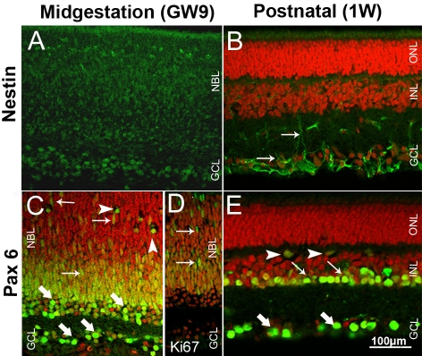
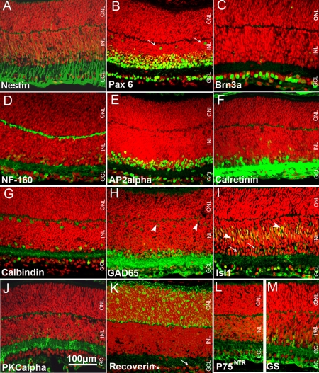
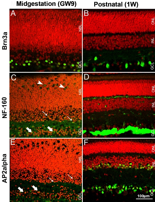
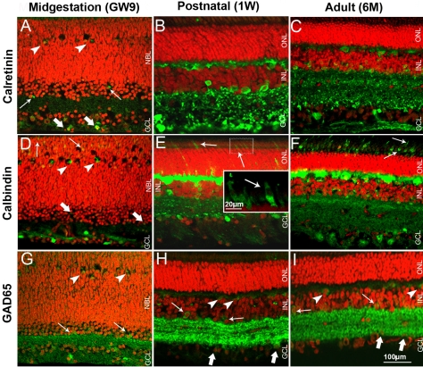
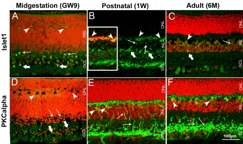
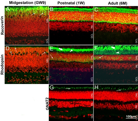
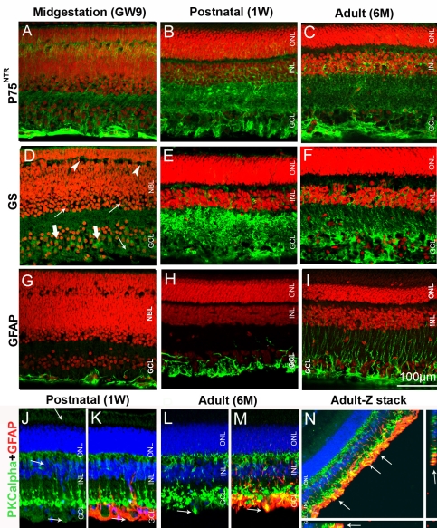
Purpose: The pig eye is similar to the human eye in terms of anatomy, vasculature, and photoreceptor distribution, and therefore provides an attractive animal model for research into retinal disease. The purpose of this study was to characterize retinal histology in the developing and mature pig retina using antibodies to well established retinal cell markers commonly used in rodents.
Methods: Eyes were enucleated from fetuses in the 9th week of gestation, 1 week old piglets and 6 months old adult animals. Eyeglobes were fixed and cryosectioned. A panel of antibodies to well established retinal markers was employed for immunohistochemistry. Fluorescently labeled secondary antibodies were used for signal detection, and images were acquired by confocal microscopy. Mouse retina at postnatal day (P) 5 was used as a reference for this study to compare progression of histogenesis. Most of the primary antibodies have previously been used on mouse tissue.
Results: Most of the studied markers were detected in midgestation pig retina, and the majority had a similar distribution in pig as in P5 mouse retina. However, rhodopsin immunolabeling was detected in pig retina at midgestation but not in P5 mouse retina. Contrary to findings in all rodents, horizontal cells were Islet1-positive and cones were calbindin-immunoreactive in pig retina, as has also been shown for the primate retina. Recoverin and rhodopsin immunolabeling revealed an increase in the length of photoreceptor segments in 6 months, compared to 1 week old animals.
Conclusions: Comparison with the published data on human retina revealed similar marker distribution and histogenesis progression in the pig and human retina, supporting the pig as a valuable animal model for studies on retinal disease and repair. Furthermore, this study provides information about the dynamics of retinal histogenesis in the pig and validates a panel of antibodies that reliably detects developing and mature retinal cell phenotypes in the pig retina.
Figures
References
-
- Chandler MJ, Smith PJ, Samuelson DA, MacKay EO. Photoreceptor density of the domestic pig retina. Vet Ophthalmol. 1999;2:179–84. - PubMed
-
- Hendrickson A, Hicks D. Distribution and density of medium- and short-wavelength selective cones in the domestic pig retina. Exp Eye Res. 2002;74:435–44. - PubMed
-
- Petters RM, Alexander CA, Wells KD, Collins EB, Sommer JR, Blanton MR, Rojas G, Hao Y, Flowers WL, Banin E, Cideciyan AV, Jacobson SG, Wong F. Genetically engineered large animal model for studying cone photoreceptor survival and degeneration in retinitis pigmentosa. Nat Biotechnol. 1997;15:965–70. - PubMed
-
- Ruiz-Ederra J, García M, Hernández M, Urcola H, Hernández-Barbáchano E, Araiz J, Vecino E. The pig eye as a novel model of glaucoma. Exp Eye Res. 2005;81:561–9. - PubMed
Publication types
MeSH terms
Substances
LinkOut - more resources
Full Text Sources
Miscellaneous