Contemporary surgery for obstructive sleep apnea syndrome
- PMID: 19784401
- PMCID: PMC2751873
- DOI: 10.3342/ceo.2009.2.3.107
Contemporary surgery for obstructive sleep apnea syndrome
Abstract
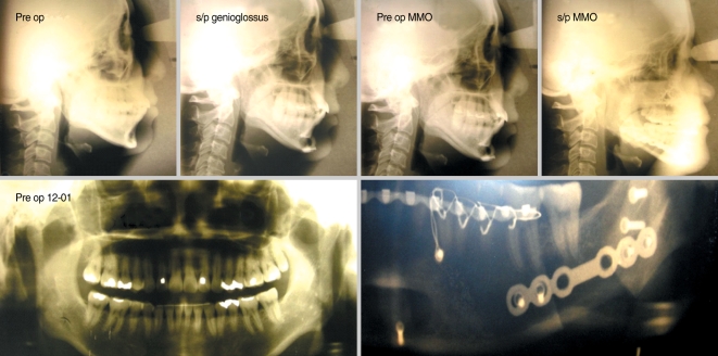
Surgical treatment of obstructive sleep apnea syndrome (OSAS) has been available in some form for greater than three decades. Early management for airway obstruction during sleep relied on tracheotomy which although life saving was not well accepted by patients. In the early eighties two new forms of treatment for OSAS were developed. Surgically a technique described as a uvulopalatopharyngoplasty (UPPP) was used to treat the retropalatal region for snoring and sleep apnea. Concurrently sleep medicine developed a nasal continuous positive airway pressure (CPAP) device to manage nocturnal airway obstruction. Both of these measures were used to expand and stabilize the pharyngeal airway space during sleep. The goal for each technique was to limit or alleviate OSAS. Almost 30 yr later these two treatment modalities continue to be the mainstay of contemporary treatment. As expected, CPAP device technology improved over time along with durable goods. Surgery followed suit and additional techniques were developed to treat soft and bony structures of the entire upper airway (nose, palate and tongue base). This review will only focus on the contemporary surgical methods that have demonstrated relatively consistent positive clinical outcomes. Not all surgical and medical treatment modalities are successful or even partially successful for every patient. Advances in the treatment of OSAS are hindered by the fact that the primary etiology is still unknown. However, both medicine and surgery continue to improve diagnostic and treatment methods. Methods of diagnosis as well as treatment regimens should always include both medical and surgical collaborations so the health and quality of life of our patients can best be served.
Keywords: Airway reconstruction; Contemporary surgery; Obstructive sleep apnea; Powell-Riley protocol.
Conflict of interest statement
I have no financial disclosure or conflict of interest with any person or company.
Figures
References
-
- Young T, Palta M, Dempsey J, Skatrud J, Weber S, Badr S. The occurrence of sleep-disordered breathing among middle-aged adults. N Engl J Med. 1993 Apr;328(17):1230–1235. - PubMed
-
- Kuhlo W, Doll E, Franck MC. Successful management of Pickwickian syndrome using long-term tracheostomy. Dtsch Med Wochenschr. 1969 Jun 13;94(24):1286–1290. - PubMed
-
- Powell N, Riley R. A surgical protocol for sleep disordered berathing. Oral Maxillofac Surg Clin North Am. 1995 Aug;7(3):345–356.
-
- Sher AE, Schechtman KB, Piccirillo JF. The efficacy of surgical modifications of the upper airway in adults with obstructive sleep apnea syndrome. Sleep. 1996 Feb;19(2):156–177. - PubMed
-
- Bachar G, Feinmesser R, Shpitzer T, Yaniv E, Nageris B, Eidelman L. Laryngeal and hypopharyngeal obstruction in sleep disordered breathing patients, evaluated by sleep endoscopy. Eur Arch Otorhinolaryngol. 2008 Nov;265(11):1397–1402. - PubMed
LinkOut - more resources
Full Text Sources
Research Materials