. 2009 Dec;66(24):3967-78.
doi: 10.1007/s00018-009-0157-5. Epub 2009 Sep 27.

Doxorubicin-mediated apoptosis in glioma cells requires NFAT3

Affiliations

Doxorubicin-mediated apoptosis in glioma cells requires NFAT3

Sreelatha Gopinath et al. Cell Mol Life Sci. 2009 Dec.

Abstract

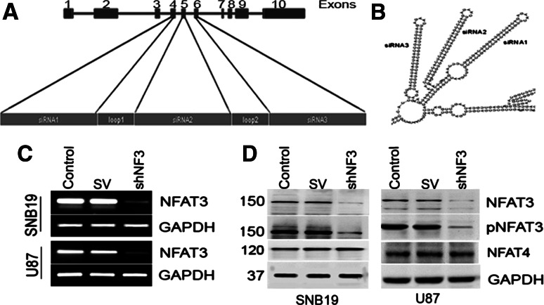
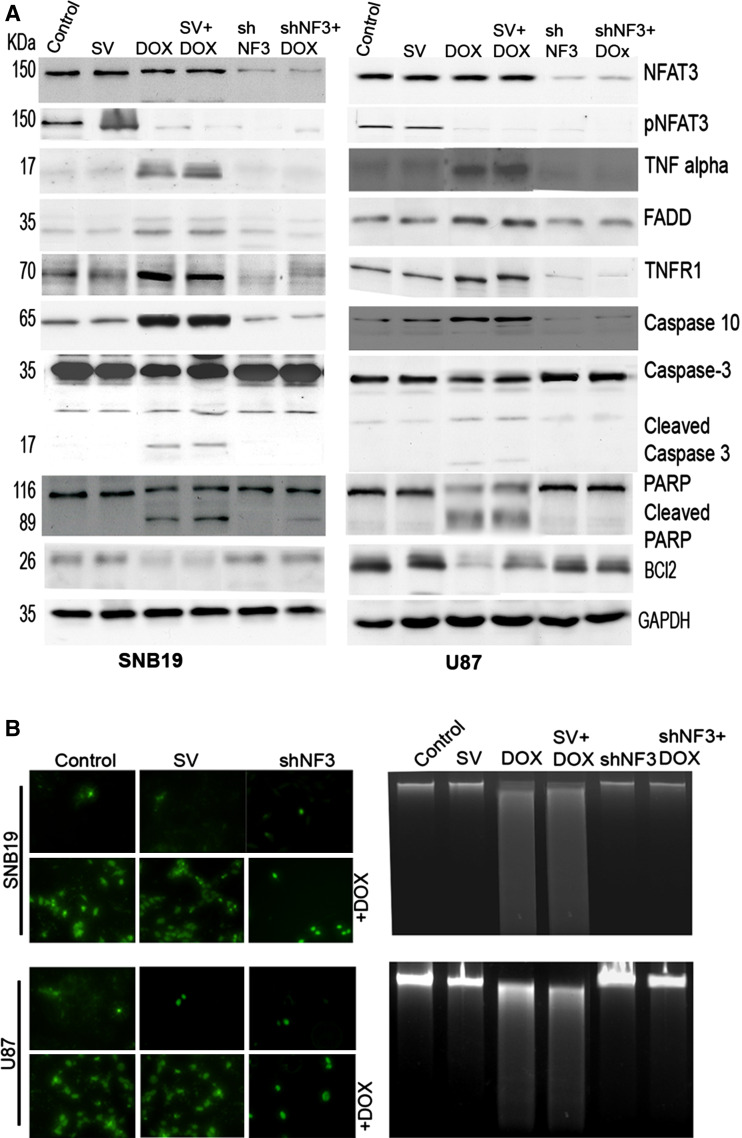
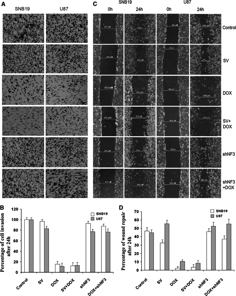
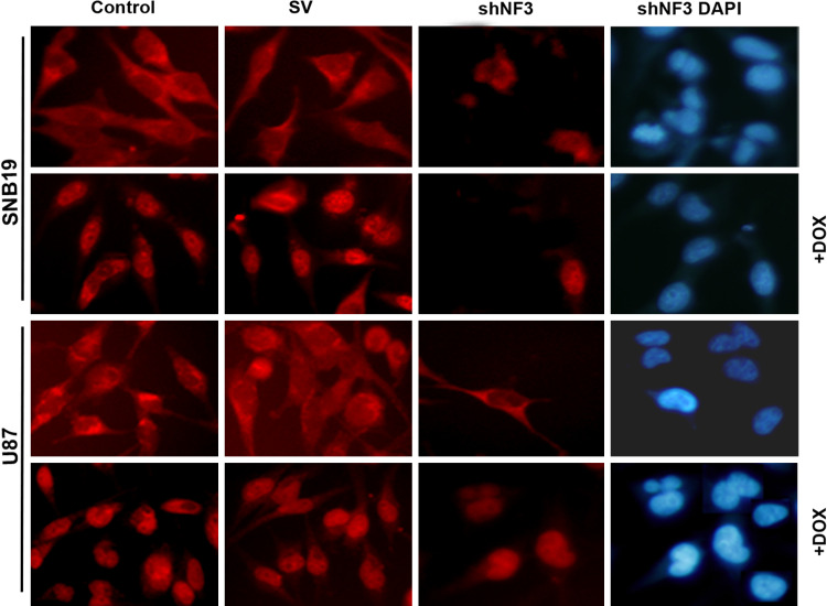
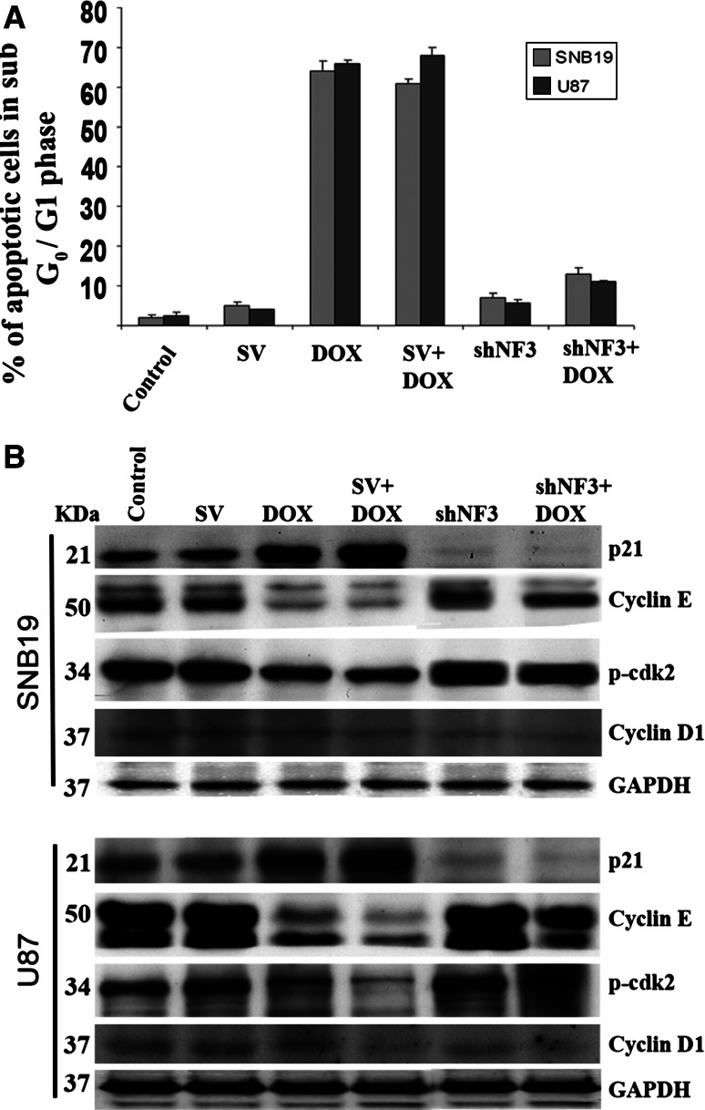
Nuclear factor of activated T cells (NFAT), a family of transcription factors, has been implicated in many cellular processes, including some cancers. Here, we characterize, for the first time, the role of NFAT3 in doxorubicin (DOX)-mediated apoptosis, migration, and invasion in SNB19 and U87 glioma cells. This study demonstrates that the specific knockdown of NFAT3 results in a dramatic inhibition of the apoptotic effect induced by DOX and favors cell survival. Inhibition of NFAT3 activation by shNFAT3 (shNF3) significantly downregulated tumor necrosis factor (TNF)-alpha induction, its receptor TNFR1, caspase 10, caspase 3, and poly (ADP-ribose) polymerase, abrogating DOX-mediated apoptosis in glioma cells. DOX treatment resulted in NFAT3 translocation to the nucleus. Similarly, shNF3 treatment in SNB19 and U87 cells reversed DOX-induced inhibition of cell migration and invasion, as determined by wound healing and matrigel invasion assays. Taken together, these results indicate that NFAT3 is a prerequisite for the induction of DOX-mediated apoptosis in glioma cells.


Figures

Fig. 1
Fig. 1
Construction of the shRNA molecule for nuclear factor of activated T cells-3 (NFAT3) and downregulation of NFAT3 in SNB19 and U87 using shNF3. a Schematic representation of the NFAT3 gene with its exons. Exons 4, 5, and 6 were targeted for the construction of short hairpin (sh) RNA for NFAT3. b Tertiary structure of the proposed shRNA showing the folds of small interfering (si) RNA 1, 2 and 3 deduced from M-fold software. c Reverse transcription (RT)-PCR analysis of glioma cells with NFAT3 primers and glyceraldehyde 3-phosphate dehydrogenase (GAPDH) primers. SV Sindbis virus. d Immunoblot analysis of cell lysates from SNB19 and U87 cells. Whole cell lysates (50-μg aliquots) from control SNB19 and U87 cells and cells transfected with SV and shNFAT3 (shNF3) for 48 h were probed with anti-NFAT3, phospho-specific anti-NFAT3 (pNFAT), and anti-NFAT4 antibodies
Fig. 2
Fig. 2
Apoptosis induced by the doxorubicin (DOX) treatment is halted in the absence of NFAT3 expression. a Cell lysates were collected from SNB19 and U87 after transfection with SV or shNF3, with or without DOX. Western blot analysis of 50 μg of total cell lysates was performed to check the expression of NFAT3, pNFAT3, Fas-associated protein with Death Domain (FADD), tumor necrosis factor alpha (TNF-α), TNF receptor 1 (TNFR1), Bcl2, caspase 3, caspase 10, and poly (ADP-ribose) polymerase (PARP). GAPDH was used as a loading control. b SNB19 and U87 cells were transfected with SV or shNF3, incubated for 16 h, treated with DOX (1 μM), and incubated for another 24 h. Apoptosis was detected using the terminal-deoxytransferase-mediated dUTP nick endlabeling (TUNEL) assay as per the manufacturer’s instructions. Cells were photographed under a light or fluorescent microscope. c Agarose gel electrophoresis pattern of DNA obtained from SNB19 and U87 cells after transfection with either SV or shNF3 and treatment with DOX, respectively
Fig. 3
Fig. 3
shNF3 treatment reversed DOX-mediated inhibition of cell invasion and wound healing. a Phase micrographs showing invading SNB19 and U87 cells. After transfection with SV or shNF3, with or without DOX, cells were trypsinized, and 2 × 105 cells were placed into matrigel-coated transwell inserts with an 8-μm pore size. After 24 h, the cells that adhered to the outer surface of the transwell (i.e., the cells which had invaded the matrigel) were fixed, stained using the Hema-3 staining kit, and counted under a light microscope. b Number of cells from ten different fields was counted for each sample, and the percentage of cells that invaded after transfection with SV or shNF3, with or without DOX, were analyzed and compared with untreated control cells. The graph represents quantification of cell invasion through the matrigel and shows the mean of ten randomly selected different fields. c SNB19 and U87 cells were cultured in six-well plate at a concentration of 2 × 106 and transfected with SV or shNF3, with or without DOX. Untreated cells were also maintained simultaneously. After the transfection and the DOX treatment were complete, a straight scratch was made in individual wells with a 200-μl pipette tip. This point was considered as the 0 h, and the width of the wound was photographed under the microscope. After 24 h, the cells were checked for wound healing and again photographed under the microscope. d Quantification of cell wound healing after cells were transfected with SV or shNF3 and/or DOX. Shown are the mean values of ten randomly selected different fields
Fig. 4
Fig. 4
DOX treatment leads to NFAT3 activation, based on its nuclear translocation. Cytosolic and nuclear presence of NFAT3 in SNB19 and U87 cells that were treated with SV or shNF3 and/or DOX. After treatment, the cells were fixed and checked for the expression of NFAT3 by the immunofluorescence assay using anti-NFAT3 primary antibody followed by fluorescein isothiocyanate-conjugated anti-rabbit secondary antibody. Cells were observed under a fluorescent microscope. DOX stimulates the NFAT3 translocation into the nucleus and the downregulation of NFAT3 resulted in the decreased expression of NFAT3 in both the cytoplasm and nucleus
Fig. 5
Fig. 5
shNF3 transfection reverses DOX-induced inhibition of cell cycle progression. a DNA content of SNB19 and U87 cells were measured by flow cytometric analysis to determine the fate of the cell cycle after transfection with SV or shNF3, with or without DOX. DOX treatment alone increased the number of apoptotic cells in the sub G0/G1 phase. Relatively fewer cells treated with shNF3 alone or with a combination of shNF3 + DOX were in the sub G0/G1 phase. The graph shows the percentage of apoptotic cells in the sub G0/G1 phase after treating SNB19 and U87 cells with SV or shNF3 and/or DOX. Values are mean ± standard deviation (SD) from three different experiments (P < 0.001). b After 48 h, total cell lysates collected from the control and transfected cells with or without DOX treatment were subjected to sodium dodecyl sulfate-polyacrylamide gel electrophoresis analysis and transferred onto a nitrocellular membrane. The membranes were probed for p21, p-cdk2, cyclin E, and cyclin D1 using the respective antibodies. GAPDH was used as the loading control

Similar articles

Cited by

References

    1. Ohgaki H, Kleihues P. Epidemiology and etiology of gliomas. Acta Neuropathol. 2005;109:93–108. doi: 10.1007/s00401-005-0991-y. - DOI - PubMed
    1. Campbell JW, Pollack IF, Martinez AJ, Shultz B. High-grade astrocytomas in children: radiologically complete resection is associated with an excellent long-term prognosis. Neurosurgery. 1996;38:258–264. doi: 10.1097/00006123-199602000-00006. - DOI - PubMed
    1. Devaux BC, O’Fallon JR, Kelly PJ. Resection, biopsy, and survival in malignant glial neoplasms. A retrospective study of clinical parameters, therapy, and outcome. J Neurosurg. 1993;78:767–775. doi: 10.3171/jns.1993.78.5.0767. - DOI - PubMed
    1. Ashkenazi A, Dixit VM. Death receptors: signaling and modulation. Science. 1998;281:1305–1308. doi: 10.1126/science.281.5381.1305. - DOI - PubMed
    1. Minotti G, Menna P, Salvatorelli E, Cairo G, Gianni L. Anthracyclines: molecular advances and pharmacologic developments in antitumor activity and cardiotoxicity. Pharmacol Rev. 2004;56:185–229. doi: 10.1124/pr.56.2.6. - DOI - PubMed

Publication types

MeSH terms