

A novel approach to tag and identify geranylgeranylated proteins
- PMID: 19784953
- PMCID: PMC2855049
- DOI: 10.1002/elps.200900259
A novel approach to tag and identify geranylgeranylated proteins
Abstract
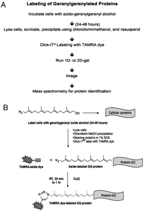
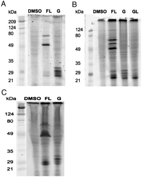
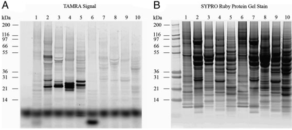
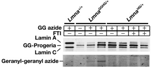
A recently developed proteomic strategy, the "GG-azide"-labeling approach, is described for the detection and proteomic analysis of geranylgeranylated proteins. This approach involves metabolic incorporation of a synthetic azido-geranylgeranyl analog and chemoselective derivatization of azido-geranylgeranyl-modified proteins by the "click" chemistry, using a tetramethylrhodamine-alkyne. The resulting conjugated proteins can be separated by 1-D or 2-D and pH fractionation, and detected by fluorescence imaging. This method is compatible with downstream LC-MS/MS analysis. Proteomic analysis of conjugated proteins by this approach identified several known geranylgeranylated proteins as well as Rap2c, a novel member of the Ras family. Furthermore, prenylation of progerin in mouse embryonic fibroblast cells was examined using this approach, demonstrating that this strategy can be used to study prenylation of specific proteins. The "GG-azide"-labeling approach provides a new tool for the detection and proteomic analysis of geranylgeranylated proteins, and it can readily be extended to other post-translational modifications.
Conflict of interest statement
The authors have declared no conflict of interest.
Figures

References
-
- Tamanoi F, Sigman DS, editors. The Enzymes. Vol. 21. San Diego: Academic Press; 2001.
-
- Zhang FL, Casey PJ. Annu. Rev. Biochem. 1996;65:241–270. - PubMed
-
- Glomset JA, Gelb MH, Farnsworth CC. Trends Biochem. Sci. 1990;15:139–142. - PubMed
-
- Cox AD, Der CJ. Curr. Opin. Cell Biol. 1992;4:1008–1106. - PubMed
Publication types
MeSH terms
Substances
Grants and funding
- GM07185/GM/NIGMS NIH HHS/United States
- R01 HL086683/HL/NHLBI NIH HHS/United States
- R01 GM066152/GM/NIGMS NIH HHS/United States
- R01 AR050200/AR/NIAMS NIH HHS/United States
- T32 GM007185/GM/NIGMS NIH HHS/United States
- HL86683/HL/NHLBI NIH HHS/United States
- CA41996/CA/NCI NIH HHS/United States
- R01 HL076839/HL/NHLBI NIH HHS/United States
- R01 AG035626/AG/NIA NIH HHS/United States
- R01 CA041996/CA/NCI NIH HHS/United States
- HL76839/HL/NHLBI NIH HHS/United States
- AR050200/AR/NIAMS NIH HHS/United States
- GM66152/GM/NIGMS NIH HHS/United States
LinkOut - more resources
Full Text Sources
