. 2009 Dec;18(12):2492-9.
doi: 10.1002/pro.254.

Stability of thioester intermediates in ubiquitin-like modifications

Affiliations

Stability of thioester intermediates in ubiquitin-like modifications

Jing Song et al. Protein Sci. 2009 Dec.

Abstract

Ubiquitin-like modifications are important mechanisms in cellular regulation, and are carried out through several steps with reaction intermediates being thioester conjugates of ubiquitin-like proteins with E1, E2, and sometimes E3. Despite their importance, a thorough characterization of the intrinsic stability of these thioester intermediates has been lacking. In this study, we investigated the intrinsic stability by using a model compound and the Ubc9 approximately SUMO-1 thioester conjugate. The Ubc9 approximately SUMO-1 thioester intermediate has a half life of approximately 3.6 h (hydrolysis rate k = 5.33 +/- 2.8 x10(-5) s(-1)), and the stability decreased slightly under denaturing conditions (k = 12.5 +/- 1.8 x 10(-5) s(-1)), indicating a moderate effect of the three-dimensional structural context on the stability of these intermediates. Binding to active and inactive E3, (RanBP2) also has only a moderate effect on the hydrolysis rate (13.8 +/- 0.8 x 10(-5) s(-1) for active E3 versus 7.38 +/- 0.7 x 10(-5) s(-1) for inactive E3). The intrinsically high stability of these intermediates suggests that unwanted thioester intermediates may be eliminated enzymatically, such as by thioesterases, to regulate their intracellular abundance and trafficking in the control of ubiquitin-like modifications.


Figures
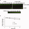
Figure 1
Figure 1
Hydrolysis of the E2∼SUMO-1 thioester. (A) Gel image of the hydrolysis experiment. SUMO was detected by Western Blot with an anti-SUMO antibody, and visualized and quantified with Li-Cor Odyssey system. Both SUMO-1 and the Ubc9∼SUMO-1 thioester are indicated. The amounts of SUMO-1 and Ubc9∼SUMO-1 thioester were calibrated using a standard curve generated from the same Western Blot with known amounts of SUMO-1 protein on the same PVDF membrane transferred from a separate SDS-PAGE (lower panel). Asterisk indicates the samples treated with reducing SDS sample buffer and boiled for 5 min before loading on the gel. (B) Plot of relative amount of Ubc9∼SUMO-1 thioester against time. Values were calculated by determining the relative intensities of the bands for the thioester.
Figure 2
Figure 2
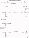
Synthesis of the model compound. Details are given in Methods.
Figure 3
Figure 3
Hydrolysis of the model compound. (A) NMR spectra recorded under native conditions showing the resonances of CH2 group of the Cys residue. The sharp peak at 3.6 ppm is from an unidentified impurity. The quartet peaks around 3.65 and 3.32ppm are from the CH2 group before hydrolysis. The upper spectrum was obtained 14 min after addition of the sample to aqueous solution, and the lower spectrum was acquired after 342 min. The boxed region was integrated to extract the hydrolysis rate. (B) Plot of the relative amount of the original compound against time, as determined by analysis of the intensities of the Cys-CH2 peak. The hydrolysis rate was deduced from linear fitting.
Figure 4
Figure 4
RanBP2 binding increased the hydrolysis of the Ubc9∼SUMO thioester. (A) Schematic of the experimental design. When hydrolysis occurs, the fluorescent SUMO-1 is released from the plate wall into the supernatant. (B) Dot blot of two-fold serial dilutions of Alexa680-SUMO-1 (1000–7.8 pg). (C) Dot blot of hydrolyzed (released) Alexa680-SUMO-1 from Ubc9∼SUMO-1 thioester immobilized on microtiter plates precoated with IR1-M-ΔSBM domain (top) or IR1-M domain (bottom). Supernatants (5 μL) taken at the indicted incubation times or supernatants (5 μL) taken from 10 min incubations in the presence of 20 mM DTT were blotted onto PVDF membrane and analyzed with an Odyssey infrared imaging system. (D) Linear regression fit of integrated intensities against Alexa680-SUMO-1 amounts for dot blot in (B). (E) Plot of relative amount (%) of released Alexa680-SUMO-1 against time, as determined by analysis of the integrated intensities of the dot blot shown in (C). The percentages of released Alexa680-SUMO-1 from microtiter plates precoated with IR1-M-ΔSBM or IR-1M were calculated relative to the amount of Alexa680-SUMO-1 released after incubation with DTT (set as 100%).

References

    1. Yeh ET, Gong L, Kamitani T. Ubiquitin-like proteins: new wines in new bottles. Gene. 2000;248:1–14. - PubMed
    1. Hershko A, Ciechanover A. The ubiquitin system. Annu Rev Biochem. 1998;67:425–479. - PubMed
    1. Varshavsky A. The ubiquitin system. Trends Biochem Sci. 1997;22:383–387. - PubMed
    1. Schwartz DC, Hochstrasser M. A superfamily of protein tags: ubiquitin, SUMO and related modifiers. Trends Biochem Sci. 2003;28:321–328. - PubMed
    1. Takada K, Hirakawa T, Yokosawa H, Okawa Y, Taguchi H, Ohkawa K. Isolation of ubiquitin-E2 (ubiquitin-conjugating enzyme) complexes from erythroleukaemia cells using immunoaffinity techniques. Biochem J. 2001;356:199–206. - PMC - PubMed

Publication types

LinkOut - more resources